Topic
Blood-brain barrier
Contents
- Anatomy of the blood-brain barrier
- Functional importance of the omega-3 DHA and its transporter Mfsd2a
- Interdependence between the intestinal and brain barriers
- Reciprocal relationship between obesity and permeability of the blood-brain barrier
- Air pollutants may breach and further compromise the blood-brain barrier
- Frequently Asked Questions
- Related Topics and Episodes
The blood-brain barrier comprises various membranes that separate the central nervous system (CNS) and the peripheral circulatory system. The purpose of this barrier is to allow the passage of nutrients and cell signals from the bloodstream to nerves and supporting cells in the CNS while excluding harmful substances. Genetic predispositions, environmental exposures, and aging may weaken the integrity of this barrier, allowing endotoxins and inflammatory immune cells to infiltrate the brain, contributing to accelerated brain aging and the risk of neurodegenerative diseases such as Alzheimer's,[1] Parkinson's,[2] and multiple sclerosis.[3]
Environmental factors shown to modulate the blood-brain barrier include:
- Omega-3 fatty acids - Omega-3s, particularly the marine-derived DHA, regulate transport across the blood-brain barrier.[4]
- Polyphenols - Polyphenols and other phytonutrients from plant-based foods increase neuroprotective metabolites at the blood-brain barrier.[5]
- Obesity - Obesity increases endotoxemia and toll-like receptor activation[6] due to perturbed intestinal permeability and hyperglycemia,[7], disrupting blood-brain barrier function.
- Aerobic exercise - Exercise reduces age-related changes to the blood-brain barrier, increasing leakiness and neurodegeneration.[8]
Anatomy of the blood-brain barrier
Blood vessels throughout the body are lined with a single layer, or sheet, of endothelial cells, a specialized type of epithelial (i.e., skin-like) cell. The endothelial sheet's luminal (inner) side interfaces directly with the bloodstream and contains transporters that exchange nutrients and waste. The endothelium's basal (outer) side at the blood-brain barrier connects mostly to pericytes, which are multipurpose cells that control the dilation and constriction of blood vessels and secrete paracrine and endocrine signals. Astroglia, star-shaped cells with many arms, connect the endothelium and pericytes of the vascular system to neurons in the CNS.[9]
This multicell structure and its many sets of connecting membranes is called a neurovascular unit and serves the purpose of nutrient and waste exchange while providing many points of regulation to prevent neuronal damage.[10] In addition to creating connections between blood vessels and neurons, astrocytes also connect blood vessels, forming a circuit for waste disposal called the glymphatic system. This highly controlled fluid movement system is responsible for waste disposal during sleep, injury, and illness because, unlike other organs, the brain has no traditional lymphatic structures.[11] Glymphatic dysfunction disturbs normal pressure in the brain and impairs waste disposal, contributing to neurodegeneration.[12]
Functional importance of the omega-3 DHA and its transporter Mfsd2a
Unsaturated fats build flexible and efficient membranes
"The composition of [fatty acids in the cell membranes] is a determining factor in membrane permeability, with saturated fats increasing rigidity and reducing permeability and unsaturated fats making membranes less compact and more fluid." Click To Tweet


MFSD2A is essential for blood-brain barrier integrity and DHA transport
Membranes throughout the body are composed of a phospholipid bilayer. Each phospholipid unit contains a hydrophilic (i.e., water-loving) head and a hydrophobic (i.e., water-averse) tail. These units arrange themselves so that the hydrophobic tails condense in the center of the membrane while the hydrophilic heads face toward the inner and outer membrane surfaces. The most common phospholipids in the body contain an amino acid or carbohydrate as part of the head connected to a glycerol molecule and two fatty acids for the tail. The composition of these fatty acids is a determining factor in membrane permeability, with saturated fats increasing rigidity and reducing permeability and unsaturated fats making membranes less compact and more fluid.[13]
The long-chain omega-3 fatty acid docosahexaenoic acid (DHA) is an example of this configuration, containing 22 carbons and six points of unsaturation where cis-oriented double carbon bonds create kinks in the phospholipid structure. Compared to phospholipids containing saturated fats, which are easily stacked and condensed, phospholipids containing DHA create a very fluid structure. Generally, more fluid membranes facilitate the movement of receptors, transporters, and other structures embedded in the membrane, improving cell metabolism.[14]
DHA blocks the entry of toxins and infection by suppressing transcytosis at the blood-brain barrier
Membrane fluidity also modulates transcytosis, the process by which molecules move from the bloodstream, through the inside of endothelial cells, and then into the surrounding tissue. An example of transcytosis is the movement of iron across the blood-brain barrier via the formation of membrane pouches called caveolae (from the Latin word for "little caves"). The body tightly controls the movement of iron in the brain to maximize enzyme efficiency without causing excessive oxidative damage. Iron is shuttled through the bloodstream by the protein transferrin.[15] When the transferrin-iron complex binds to transferrin receptors on endothelial cells, the physical shape of the receptor changes, initiating a cascade of motion. The receptor and surrounding membrane are pulled into the cell, while scaffolding proteins called caveolin coat the budding membrane and help pinch it closed.[16]
The finished vesicle resembles a sphere made of cellular membrane coated with caveolin on the outside and filled with fluid from the bloodstream. The transferrin receptor complex is located inside the membrane vesicle and is released in a regulated way inside the cell or secreted to surrounding cells needing iron. Transcytotic vesicles such as caveolae facilitate effective sorting of cellular packages, delivering cell signals to the Golgi apparatus, hormones to the endoplasmic reticulum,[17] and cellular debris to lysosomes;[18] however, this system can be hijacked by pathogens such as viruses, which use it to invade cells.[19] Therefore, the system must weigh the benefits of efficient metabolic exchange against the risk of infection and injury. Compared to endothelial cells in peripheral organs such as the lungs, which utilize caveolae and other methods of vesicle-mediated transcytosis to a large extent, endothelial cells at the blood-brain barrier severely limit transcytosis.[20]
The brain uses special transporters to enrich itself with DHA
Transcytotic vesicles often form in lipid rafts, areas of the cellular membrane enriched with saturated fats and cholesterol that stabilize the vesicle structure.[21] Therefore, blood-brain barrier cells are enriched with DHA, which has a long, irregular shape that prevents the membrane condensation needed to form caveolae. Blood-brain barrier endothelia express receptors such as Mfsd2a that transport lipids across the barrier to accumulate DHA.[22]
Findings from a 2017 study demonstrate that Mfsd2a is needed to suppress caveolae-mediated transcytosis and protect the integrity of the blood-brain barrier. The report's authors previously demonstrated that Mfsd2a, a transmembrane protein found exclusively on the endothelial cells of the blood-brain barrier, is the sole means by which phospholipid DHA passes into the brain.[22] In a follow-up experiment using mice that carried a mutation blocking Mfsd2a's capacity to transport DHA, the investigators assessed barrier function, caveolae formation, and activity in the animals' brains. Then, they compared the lipid composition of brain endothelial cells to lung epithelial cells, which lack Mfsd2a.
Endothelial cells making up the blood-brain barrier had uniquely high levels of DHA. They found that mice that lacked Mfsd2a function had reduced blood-brain barrier integrity and greater caveolae formation and activity than normal mice. In normal mice, they also found that brain endothelial cells had higher lipid concentrations than lung epithelial cells. The most abundant lipid was DHA, found in concentrations two to five times higher. These findings suggest that Mfsd2a-mediated transport of lipids, particularly DHA, impairs caveolae activity, thereby preserving blood-brain integrity.[23]
More discussion of the role of Mfsd2a, DHA, and the brain in this peer-reviewed article by Dr. Rhonda Patrick:
- Patrick, Rhonda P. "Role of phosphatidylcholine-DHA in preventing APOE4-associated Alzheimer's disease." The FASEB Journal 33.2 (2019): 1554-1564.
Interdependence between the intestinal and brain barriers
The intestinal barrier is the largest membrane interface between the body and the outside world. Like all membranous barriers, it serves the purpose of exchanging nutrients and waste, but on an exponentially larger scale than most. Also similar to other membranous barriers, the intestinal barrier loses function with age and disease. Increased intestinal permeability, also known as "leaky gut," compromises the health of the whole body by allowing the passage of pathogens and toxins from food and beverages to enter the bloodstream.[24]
Unlike the blood-brain barrier, the intestinal barrier has the added complexity of interfacing with the gut microbiota, the community of microorganisms that inhabits the digestive tract. The intestinal barrier utilizes physical, chemical, and biological strategies to maintain distance between intestinal cells and pathogens and toxins moving through the digestive tract. The intestinal epithelium contains goblet cells that secrete a layer of protective mucus that restricts the growth of bacteria and the movement of toxins. Immune cells in the intestinal epithelium secrete immune factors into the mucus layer, allowing friendly bacteria to colonize space close to the barrier, crowding out unfamiliar microbes. When this complex filtering system weakens, microbial toxins in the bloodstream increase, a condition known as endotoxemia.[25]
Intestinal hyperpermeability causes endotoxemia and accelerated brain aging
The most common endotoxin in circulation is lipopolysaccharide (LPS), a lipid and carbohydrate structure shed from the cell walls of Gram-negative bacteria such as Escherichia coli. People with intestinal disorders, such as celiac disease[26] and inflammatory bowel disease,[27] experience chronic barrier disruption and endotoxemia. This phenomenon also occurs in conditions such as obesity and type 2 diabetes, characterized by metabolic endotoxemia, a chronic two- to threefold elevation in endotoxins that increases whole-body inflammation and contributes to insulin resistance and weight gain.[28] Many diseases involving the brain also share chronic mild endotoxemia as a feature, including neurodevelopmental disorders such as autism spectrum disorder[29], neuropsychiatric diseases such as depression[30], and neurodegenerative diseases such as Alzheimer's disease[31] and Parkinson's disease.[32] This wide-ranging list of conditions involving perturbation of the gut-brain axis continues to grow.
Research investigating the role of the gut-brain axis in Alzheimer's disease provides a greater understanding of how endotoxemia affects the blood-brain barrier and better brain health in the context of aging. The body of available research demonstrates that alterations in the gut microbiota composition reduce gut barrier integrity and expose the immune system to pathogen-associated molecular patterns such as LPS. Toll-like receptor (TLR)4s on monocytes and endothelial cells intercept LPS,[33] causing systemic inflammation that increases blood-brain barrier permeability and promotes neurodegeneration.
Membrane permeability is required to recruit immune cells and nutrients to areas of damage during an infection or after an injury. Damage-associated molecular patterns (DAMPs) such as high-mobility group protein (HMG)-1, which organizes chromatin and controls DNA transcription, also bind to immune receptors that stimulate inflammation. In the case of DAMP receptor activation, cells of the blood-brain barrier recruit phagocytic (i.e., cell-eating) immune cells such as monocytes to clear cellular debris and stimulate repair. DAMPs such as HMG-1 bind to TLR4 and receptors for advanced glycation end products (RAGE) on cells at the blood-brain barrier to induce an immune response that initially inhibits the deposition of amyloid-beta, a key feature of Alzheimer's disease progression. With chronic activation, the immune system's response accelerates neurodegeneration.[34] Future investigations should expand on this work by discovering therapies that ameliorate DAMP-induced inflammation to treat neurodegenerative disorders.
Reciprocal relationship between obesity and permeability of the blood-brain barrier
In obesity, insulin-sensitive tissues such as the liver, adipose tissue, and skeletal muscle are overwhelmed by a chronic intake of excess calories, usually from high-glycemic index foods. These cells reduce their responsiveness to insulin to protect themselves from the oxidative stress caused by excess caloric intake. As insulin resistance worsens, blood sugar levels become chronically elevated, leading to long-term remodeling of the blood-brain barrier. Changes include the thickening of membranes, a reduction of blood flow, and increased permeability.[35]
Consequences of hyperglycemia
"AGEs directly reduce the expression of the tight-junction proteins in pericytes, permeabilizing the blood-brain barrier and increasing the production of pro-inflammatory cytokines." Click To Tweet
One reason for this increased permeability is the production of advanced glycation end products (AGEs), which are lipid and protein structures that have been glycated, meaning chemically bonded to glucose. AGEs directly reduce the expression of the tight-junction proteins in pericytes, permeabilizing the blood-brain barrier and increasing the production of pro-inflammatory cytokines.[36] Obesity further injures the blood-brain barrier by reducing the expression of cytoskeletal proteins necessary for supporting the barrier, transport proteins that facilitate nutrient and waste exchange, and functional proteins such as enzymes, chaperones, and transcription factors. In turn, the blood-brain barrier's capacity to transport appetite-suppressing hormones such as leptin into the CNS diminishes. The absence of these satiating signals drives overeating, promoting further weight gain.[37]
Air pollutants may breach and further compromise the blood-brain barrier
"Alarmingly, researchers found early signs of Alzheimer's disease pathology in the brains of children and young adults exposed to high levels of air pollution in Mexico City." Click To Tweet
Mounting evidence suggests that air pollution is harmful to brain health. Pollutants like ozone, sulfur dioxide, nitrogen oxides, and fine particulate matter from traffic, industry, and wildfires can enter the brain's olfactory bulb through inhalation or reach the brain through intestinal and blood-brain barriers when ingested. Once inside the brain, contaminants may trigger immune cells to release cytokines, which drive neuroinflammation. Exposure to airborne pollutants may increase the risk of neurodegenerative diseases, including Alzheimer's [38] and Parkinson's.[39] Preclinical studies suggest that particulate matter air pollution can interact with and disrupt gut bacteria.[40]
Populations most vulnerable to the damaging effects of air pollution include children and older adults.[41] The structure of the blood-brain barrier shifts across the lifecycle from a developmental state in neonates to a weakened state in old age, potentially allowing contaminants to infiltrate the brain during these stages more readily.[42] Similarly, one in four people carry an APOE4 gene variant, which increases their risk for Alzheimer's disease and may render them more susceptible to air pollution-induced neuroinflammatory damage.[43]
Alarmingly, researchers found early signs of Alzheimer's disease pathology in the brains of children and young adults exposed to high levels of air pollution in Mexico City, an area known for having high levels of air pollution. Autopsy findings revealed elevated levels of amyloid and tau proteins in 202 out of 203 young brains, including an 11-month-old baby.[44]
Preclinical evidence suggests a role for toll-like receptor 4 (TLR4) activation in air-pollution-induced neuroinflammation.[45] Toll-like receptors detect the presence of bacterial components and signal the body to secrete inflammatory cytokines. Airborne particulate matter is sometimes associated with lipopolysaccharides derived from Gram-negative bacteria (PM-LPS) and can activate TLR4 receptors and exacerbate inflammation, particularly in the lungs.[46]
Frequently Asked Questions
Q: What lifestyle factors prevent or ameliorate blood-brain leakage?
A: Many lifestyle factors help maintain blood-brain barrier integrity, including:
Maintaining low visceral fat: Visceral fat, which accumulates around the abdominal organs, promotes inflammation and increases the risk of metabolic syndrome. Elevated levels of visceral fat are linked to a compromised blood-brain barrier.
- High omega-3 fatty acid intake: Omega-3 fatty acids help maintain the integrity of the blood-brain barrier and reduce inflammation. Learn more about omega-3s and brain health in our comprehensive overview article.
Omega-3 Index validation: The Omega-3 Index is a biomarker of omega-3 fatty acids in red blood cells, providing a more accurate assessment of omega-3 status and informing dietary and supplemental intake. Learn more in our article.
Aerobic exercise: Aerobic exercise helps maintain healthy blood pressure. Having high blood pressure can compromise the blood-brain barrier, allowing harmful substances, such as amyloid proteins, to enter the brain. Learn more in this clip.
Related Topics and Episodes
Topic Pages
Intestinal permeability - Intestinal permeability is a source of stress for the blood-brain barrier that increases permeability, injuring neurons and promoting disease.
Toll-like receptors - Toll-like receptors are the principal inducers of innate immunity and participate directly in the loosening of the blood-brain barrier in the setting of endotoxemia.
Aerobic exercise - Exercise reduces age-related changes to the blood-brain barrier that increases leakiness and neurodegeneration.
Polyphenols - Polyphenols and other phytonutrients from plant foods increase neuroprotective metabolites at the blood-brain barrier.
Episodes & Clips
- ^ Deane, Rashid; Zlokovic, Berislav (2007). Role Of The Blood-Brain Barrier In The Pathogenesis Of Alzheimers Disease Current Alzheimer Research 4, 2.
- ^ Bartels, A. L.; Willemsen, A. T. M.; Kortekaas, R.; De Jong, B. M.; De Vries, R.; De Klerk, O., et al. (2008). Decreased Blood–Brain Barrier P-glycoprotein Function In The Progression Of Parkinson’s Disease, PSP And MSA Journal Of Neural Transmission 115, 7.
- ^ Minagar, Alireza; Alexander, J Steven (2003). Blood-brain Barrier Disruption In Multiple Sclerosis Multiple Sclerosis Journal 9, 6.
- ^ Randall, Elizabeth C.; Lopez, Begoña G. C.; Regan, Michael S.; Andreone, Benjamin J.; Gu, Chenghua; Agar, Jeffrey N., et al. (2020). Spatial Distribution Of Transcytosis Relevant Phospholipids In Response To Omega-3 Dietary Deprivation ACS Chemical Biology 16, 1.
- ^ Kirk, Riley D.; Seeram, Navindra P.; DaSilva, Nicholas; Ma, Hang; Bertin, Matthew; Johnson, Shelby L. (2019). Polyphenol Microbial Metabolites Exhibit Gut And Blood–Brain Barrier Permeability And Protect Murine Microglia Against LPS-Induced Inflammation Metabolites 9, 4.
- ^ Haruwaka, Koichiro; Ikegami, Ako; Ohno, Nobuhiko; Matsumoto, Mami; Kato, Daisuke; Ono, Riho, et al. (2019). Dual Microglia Effects On Blood Brain Barrier Permeability Induced By Systemic Inflammation Nature Communications 10, 1.
- ^ Lacoste, Baptiste; Dyken, Peter Van (2018). Impact Of Metabolic Syndrome On Neuroinflammation And The Blood–Brain Barrier Frontiers In Neuroscience 12, .
- ^ Graham, Leah C.; Simeone, Stephen N.; Radell, Jake E.; Funkhouser, W. Keith; Howell, Megan C.; Howell, Gareth R., et al. (2015). APOE Stabilization By Exercise Prevents Aging Neurovascular Dysfunction And Complement Induction PLOS Biology 13, 10.
- ^ Nelson, Amy R.; Betsholtz, Christer; Zlokovic, Berislav V.; Zhao, Zhen (2015). Establishment And Dysfunction Of The Blood-Brain Barrier Cell 163, 5.
- ^ Iadecola C (2017). The Neurovascular Unit Coming of Age: A Journey through Neurovascular Coupling in Health and Disease. Neuron 96, 1.
- ^ Plog, Benjamin A.; Nedergaard, Maiken (2018). The Glymphatic System In Central Nervous System Health And Disease: Past, Present, And Future Annual Review Of Pathology: Mechanisms Of Disease 13, 1.
- ^ Karimy, Jason K.; Kundishora, Adam J.; Mestre, Humberto; Cerci, H. Mert; Matouk, Charles; Alper, Seth L., et al. (2020). Glymphatic System Impairment In Alzheimer’s Disease And Idiopathic Normal Pressure Hydrocephalus Trends In Molecular Medicine 26, 3.
- ^ Cazzola, Roberta; Rondanelli, Mariangela; Russo-Volpe, Samantha; Ferrari, Ettore; Cestaro, Benvenuto (2004). Decreased Membrane Fluidity And Altered Susceptibility To Peroxidation And Lipid Composition In Overweight And Obese Female Erythrocytes Journal Of Lipid Research 45, 10.
- ^ Yang, Xiaoguang; Sheng, Wenwen; Sun, Grace Y.; Lee, James C.-M. (2011). Effects Of Fatty Acid Unsaturation Numbers On Membrane Fluidity And Α-Secretase-Dependent Amyloid Precursor Protein Processing Neurochemistry International 58, 3.
- ^ Aoki, Takeo; Hagiwara, Haruo; Matsuzaki, Toshiyuki; Suzuki, Takeshi; Takata, Kuniaki (2007). Internalization Of Caveolae And Their Relationship With Endosomes In Cultured Human And Mouse Endothelial Cells Anatomical Science International 82, 2.
- ^ Lian, Xiaoming; Kaßmann, Mario; Daumke, Oliver; Gollasch, Maik; Matthaeus, Claudia (2019). Pathophysiological Role Of Caveolae In Hypertension Frontiers In Medicine 6, .
- ^ Le, Phuong U.; Nabi, Ivan Robert (2003). Distinct Caveolae-Mediated Endocytic Pathways Target The Golgi Apparatus And The Endoplasmic Reticulum Journal Of Cell Science 116, 6.
- ^ Kiss, Anna L.; Botos, Erzsébet (2009). Endocytosisviacaveolae: Alternative Pathway With Distinct Cellular Compartments To Avoid Lysosomal Degradation? Journal Of Cellular And Molecular Medicine 13, 7.
- ^ Xing, Yifan; Wen, Zeyu; Gao, Wei; Lin, Zhekai; Zhong, Jin; Jiu, Yaming (2020). Multifaceted Functions Of Host Cell Caveolae/Caveolin-1 In Virus Infections Viruses 12, 5.
- ^ Oh, Phil; Borgström, Per; Witkiewicz, Halina; Li, Yan; Borgström, Bengt J; Iwata, Koji, et al. (2007). Live Dynamic Imaging Of Caveolae Pumping Targeted Antibody Rapidly And Specifically Across Endothelium In The Lung Nature Biotechnology 25, 3.
- ^ Galbiati F; Razani B; Lisanti MP (2001). Emerging themes in lipid rafts and caveolae. Cell 106, 4.
- ^ a b Ben-Zvi, Ayal; Lacoste, Baptiste; Kur, Esther; Andreone, Benjamin J.; Yan, Han; Gu, Chenghua, et al. (2014). Mfsd2a Is Critical For The Formation And Function Of The Blood–Brain Barrier Nature 509, 7501.
- ^ Andreone, Benjamin J.; Chow, Brian Wai; Tata, Aleksandra; Lacoste, Baptiste; Ben-Zvi, Ayal; Bullock, Kevin, et al. (2017). Blood-Brain Barrier Permeability Is Regulated By Lipid Transport-Dependent Suppression Of Caveolae-Mediated Transcytosis Neuron 94, 3.
- ^ Farhadi, Ashkan; Banan, Ali; Fields, Jeremy; Keshavarzian, Ali (2003). Intestinal Barrier: An Interface Between Health And Disease Journal Of Gastroenterology And Hepatology 18, 5.
- ^ Wells, Jerry M.; Brummer, Robert J.; MacDonald, Thomas T.; Troost, Freddy; Theodorou, Vassilia; Dekker, Jan, et al. (2017). Homeostasis Of The Gut Barrier And Potential Biomarkers American Journal Of Physiology-Gastrointestinal And Liver Physiology 312, 3.
- ^ 10.5114/ceji.2021.107012
- ^ Dejban, Pegah; Nikravangolsefid, Nasrin; Chamanara, Mohsen; Dehpour, Ahmadreza; Rashidian, Amir (2020). The Role Of Medicinal Products In The Treatment Of Inflammatory Bowel Diseases ( IBD ) Through Inhibition Of TLR4 / NF‐kappaB Pathway Phytotherapy Research 35, 2.
- ^ Costa, Jorge De Assis; Alfenas, Rita De Cássia Gonçalves; Gomes, Júnia Maria Geraldo (2017). Metabolic Endotoxemia And Diabetes Mellitus: A Systematic Review Metabolism 68, .
- ^ Jyonouchi, Harumi; Geng, Lee; Ruby, Agnes; Zimmerman-Bier, Barbie (2005). Dysregulated Innate Immune Responses In Young Children With Autism Spectrum Disorders: Their Relationship To Gastrointestinal Symptoms And Dietary Intervention Neuropsychobiology 51, 2.
- ^ Lekander, Mats; Schedlowski, Manfred; Engler, Harald; Benson, Sven; Lasselin, Julie (2020). Sick For Science: Experimental Endotoxemia As A Translational Tool To Develop And Test New Therapies For Inflammation-Associated Depression Molecular Psychiatry 26, 8.
- ^ André, Perrine; Samieri, Cécilia; Buisson, Charline; Dartigues, Jean-François; Helmer, Catherine; Laugerette, Fabienne, et al. (2019). Lipopolysaccharide-Binding Protein, Soluble CD14, And The Long-Term Risk Of Alzheimer’s Disease: A Nested Case-Control Pilot Study Of Older Community Dwellers From The Three-City Cohort Journal Of Alzheimer's Disease 71, 3.
- ^ Jackson, Aeja; Forsyth, Christopher B.; Shaikh, Maliha; Voigt, Robin M.; Engen, Phillip A.; Ramirez, Vivian, et al. (2019). Diet In Parkinson's Disease: Critical Role For The Microbiome Frontiers In Neurology 10, .
- ^ Kowalski, Karol; Mulak, Agata (2019). Brain-Gut-Microbiota Axis In Alzheimer’s Disease Journal Of Neurogastroenterology And Motility 25, 1.
- ^ Aamir, Khurram; Shaikh, Mohd. Farooq; Othman, Iekhsan; Paudel, Yam Nath; Angelopoulou, Efthalia; Piperi, Christina (2020). Impact Of HMGB1, RAGE, And TLR4 In Alzheimer’s Disease (AD): From Risk Factors To Therapeutic Targeting Cells 9, 2.
- ^ Prasad S; Sajja RK; Naik P; Cucullo L (2014). Diabetes Mellitus and Blood-Brain Barrier Dysfunction: An Overview. J Pharmacovigil 2, 2.
- ^ Shimizu, Fumitaka; Sano, Yasuteru; Tominaga, Osamu; Maeda, Toshihiko; Abe, Masa-aki; Kanda, Takashi (2013). Advanced Glycation End-Products Disrupt The Blood–Brain Barrier By Stimulating The Release Of Transforming Growth Factor–Β By Pericytes And Vascular Endothelial Growth Factor And Matrix Metalloproteinase–2 By Endothelial Cells In Vitro Neurobiology Of Aging 34, 7.
- ^ Ouyang, Suidong; Hsuchou, Hung; Kastin, Abba J; Wang, Yuping; Yu, Chuanhui; Pan, Weihong (2013). Diet-Induced Obesity Suppresses Expression Of Many Proteins At The Blood–Brain Barrier British Journal Of Pharmacology 34, 1.
- ^ Nakayama, Shoji F.; Tsai, Chon-Haw; Sun, Xian-Liang; Ma, Chaochen; Hwang, Bing-Fang; Tsai, Tsung-Lin, et al. (2019). Fine Particulate Matter Is A Potential Determinant Of Alzheimer's Disease: A Systemic Review And Meta-Analysis Environmental Research 177, .
- ^ Goldman, Sam (2014). Environmental Toxins And Parkinson's Disease Annual Review Of Pharmacology And Toxicology 54, 1.
- ^ Engen, Phillip; Mutlu, Ece A.; Comba, Işın Y.; Cho, Takugo; Yazıcı, Cemal; Soberanes, Saul, et al. (2018). Inhalational Exposure To Particulate Matter Air Pollution Alters The Composition Of The Gut Microbiome Environmental Pollution 240, .
- ^ Schins, Roel P.F.; Wahle, Tina; Campbell, Arezoo; Westerink, Remco H.S.; Tran, Lang; Johnston, Helinor, et al. (2016). Neurodegenerative And Neurological Disorders By Small Inhaled Particles NeuroToxicology 56, .
- ^ Dhaliwal, Satvinder S; Mamo, John C.L; Giles, Corey; Takechi, Ryusuke; Jackaman, Connie; Lam, Virginie, et al. (2015). Blood–brain Barrier Dysfunction Developed During Normal Aging Is Associated With Inflammation And Loss Of Tight Junctions But Not With Leukocyte Recruitment Immunity & Ageing 12, 1.
- ^ Wang, X; Driscoll, I; Woodward, N; Saffari, A; Reyes, J; Serre, M L, et al. (2017). Particulate Air Pollutants, APOE Alleles And Their Contributions To Cognitive Impairment In Older Women And To Amyloidogenesis In Experimental Models Translational Psychiatry 7, 1.
- ^ Torres, Ricardo; Calderón-Garcidueñas, Lilian; Gónzalez-Maciel, Angélica; Reynoso-Robles, Rafael; Delgado-Chávez, Ricardo; Mukherjee, Partha S., et al. (2018). Hallmarks Of Alzheimer Disease Are Evolving Relentlessly In Metropolitan Mexico City Infants, Children And Young Adults. APOE4 Carriers Have Higher Suicide Risk And Higher Odds Of Reaching NFT Stage V At ≤ 40 Years Of Age Environmental Research 164, .
- ^ Woodward, Nicholas C.; Levine, Morgan C.; Shirmohammadi, Farimah; Saffari, Arian; Morgan, Todd E.; Finch, Caleb E., et al. (2017). Toll-like Receptor 4 In Glial Inflammatory Responses To Air Pollution In Vitro And In Vivo Journal Of Neuroinflammation 14, 1.
- ^ Fonceca, Angela M.; Bozanich, Elizabeth M.; Sutanto, Erika N.; McNamara, Paul S.; Knight, Darryl A.; Turner, Debra J., et al. (2018). Accumulation Mode Particles And LPS Exposure Induce TLR-4 Dependent And Independent Inflammatory Responses In The Lung Respiratory Research 19, 1.
Episodes related to Blood-brain barrier
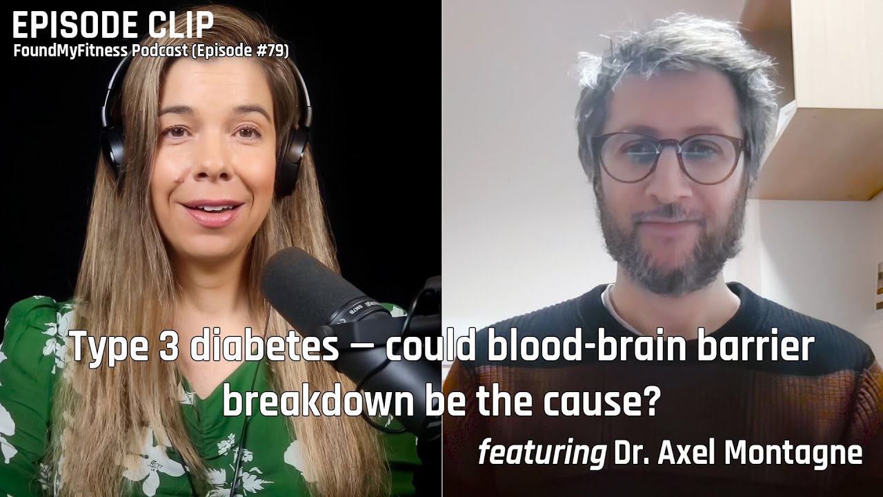
"Type 3 diabetes" — could blood-brain barrier breakdown be the cause? | Axel Montagne, Ph.D.

Topics related to Blood-brain barrier
-
Salmon roe
Salmon roe, the internal egg mass found in female salmon, is rich in protein, vitamins, and the omega-3 fatty acids EPA and DHA.
-
Brain-derived neurotrophic factor (BDNF)
BDNF is a growth factor known for its influence on neuronal health and for its role in mediating the beneficial cognitive effects associated with exercise.
-
Omega-3 fatty acids
Omega-3 fatty acids play critical roles in human health and may be beneficial in ameliorating symptoms associated with chronic health conditions and in combating aging-related diseases.
-
Omega-3 Index
The Omega-3 Index measures the amount of EPA + DHA as a percentage of total fatty acids in red blood cell membranes.
-
Polyphenol-Rich Diets and Brain Aging
Polphenol-rich diets promote better glucose regulation can help slow brain aging, preserve hippocampal function, and reduce cognitive decline over time.
-
Choline
Choline is an essential nutrient critical for various bodily functions, including brain development, liver health, and muscle function.