Cognition
Episodes
When is the best time to exercise? It depends on your goal. Because your body...
In this clip, Dr. Rhonda Patrick explores lifestyle and genetic factors affecting dementia risk, and how exercise, sleep, and nutrition protect...

In this clip, Dr. Rhonda Patrick shares her top superfoods, omega-3 benefits for mood and longevity, and key nutrients that protect against...
-
When is the best time to exercise? It depends on your goal. Because your body...
-
In this clip, Dr. Rhonda Patrick explores lifestyle and genetic factors affecting dementia risk, and how exercise, sleep, and nutrition protect...
-
In this clip, Dr. Rhonda Patrick shares her top superfoods, omega-3 benefits for mood and longevity, and key nutrients that protect against...
-
In this clip, Dr. Rhonda Patrick explains why she's increased her creatine dose, how that supports brain function during...
-
In this clip, Dr. Rhonda Patrick shares why creatine supports brain health, who benefits most, and the dosing strategies she personally...
-
In this clip, Dr. Rhonda Patrick explores vigorous exercise, lactate's cognitive benefits, and why intensity often outperforms volume for brain...
-
In this clip, Dr. Rhonda Patrick discusses how sleep loss affects glucose metabolism, cognitive performance, and how vigorous exercise intensity can...
-
In this clip, Dr. Rhonda Patrick explains brain fog, how meal timing and "exercise snacks" affect cognition, and how omega-3s combat...
-
In this clip from Dr. Rhonda Patrick's appearance on the Modern Wisdom podcast, she details seven science-based methods for improving cognition.
-
In this Aliquot, Dr. Andrew Huberman, Brady Holmer, and I explore the science behind cognitive enhancers and how HIIT boosts...
-
In this clip from the Rich Roll Podcast, Dr. Rhonda Patrick explains how exercise-driven brain changes support learning, memory, and mental health.
-
In this clip, Dr. Rhonda Patrick discusses nicotine's cognitive effects, risks to health, and safer alternatives for enhancing mental performance.
-
Dr. Darren Candow and Dr. Rhonda Patrick discuss the science of creatine for muscle performance and brain health.
-
In this Aliquot, Drs. Matt Walker, Satchin Panda, and Andrew Huberman explore how to harness the benefits of natural light...
-
In this clip, Drs. Stuart Phillips and Rhonda Patrick discuss creatine's benefits for muscle growth, brain health, supplementation tips, and safety.
-
In this clip, Dr. Rhonda Patrick highlights the health benefits of cocoa flavanols for circulation, cognition, and skin, plus her dosing insights.
-
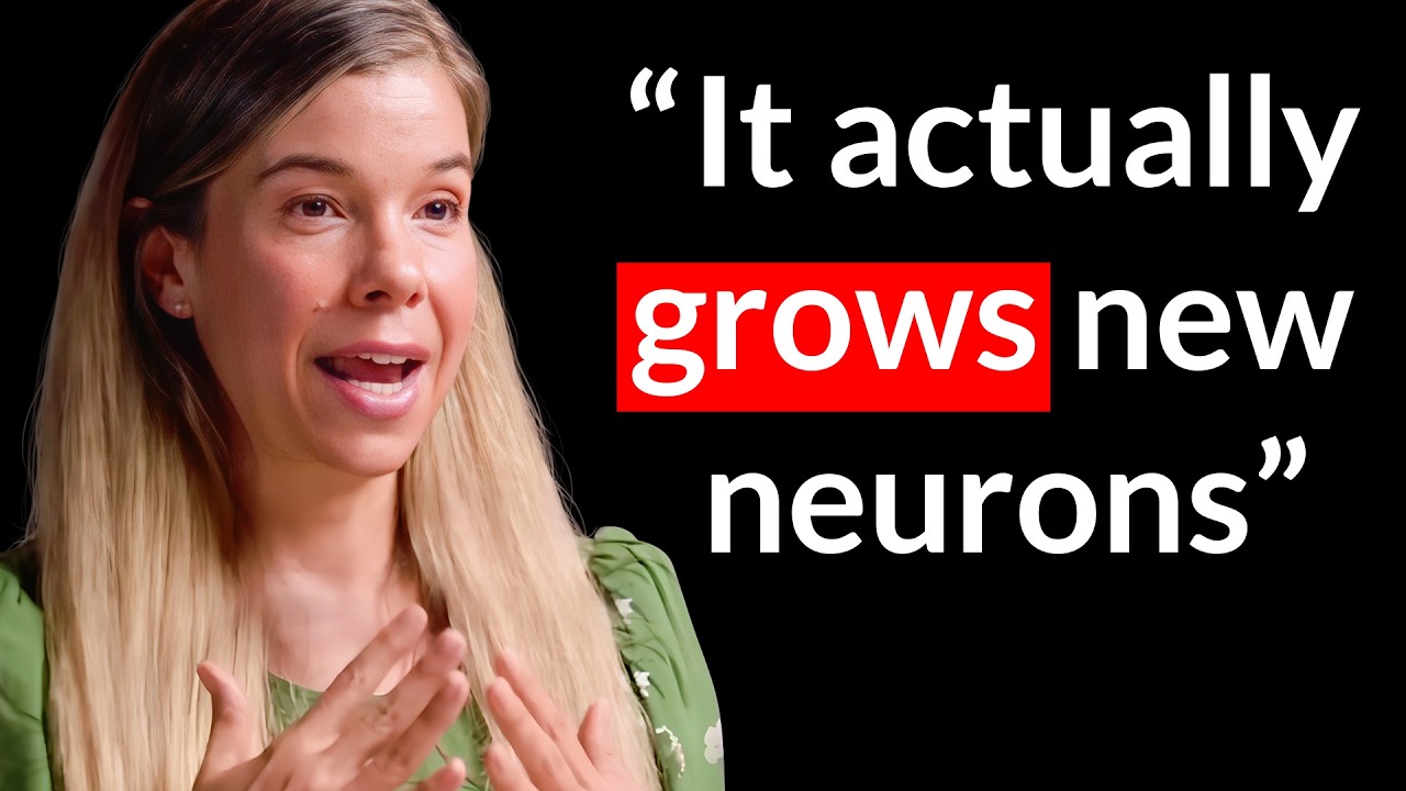
In this clip, Dr. Rhonda Patrick explains how exercise boosts memory, cognition, and neurogenesis through serotonin and post-learning activity.
-
Lactate often gets a bad rap due to its close association with lactic acid. But lactate plays a distinctly different and often-underappreciated role...
-
In this clip, Dr. Andrew Huberman discusses the power of effort, dopamine in motivation, and the mindset needed for effective learning and growth.
-
In this clip, Dr. Rhonda Patrick discusses lion's mane's potential brain benefits, its effect on mental health, and her preferred brain-enhancing supplement.
-
In this clip, Dr. Andrew Huberman discusses dopamine's role in motivation and strategies for maintaining stable levels.
-
In this clip, Dr. Rhonda Patrick discusses alcohol's effects on brain health, dementia risk, genetics, and whether resveratrol offers protective benefits.
-
In this clip, Dr. Andrew Huberman discusses using negative visualization, discomfort, and cold exposure to overcome procrastination and boost motivation.
-
In this clip, Dr. Andrew Huberman discusses how caffeine and other stimulants impact the release of dopamine in the brain.
-
In this clip, Dr. Rhonda Patrick discusses magnesium's benefits for brain health, including its potential to effectively manage migraines.
-
In this clip, Dr. Martin Gibala delves into the cognitive advantages of engaging in high-intensity exercise.
-
Meditation is a form of mental training geared toward improving aspects of a person's core neurocognitive function, such as the regulation of attention and e...
-
In this clip, Dr. George Church explains how sleeping and dreaming can inspire scientific ideas and artistic vision.
-
Chocolate and its principal component, cocoa, are popular topics – no doubt because they taste so good. But a wide range of beneficial health effects have be...
-
Sleep affects aspects of our mental and cognitive health. Not getting enough sleep triggers the onset of a "loneliness phenotype," driving people who are sle...
-
Keeping our brains sharp throughout the lifespan is essential for optimal performance. Lifestyle behaviors such as exercise, dietary modification, and sauna ...
Topic Pages
News & Publications
-
A single 14-gram dose of creatine helped preserve cognitive performance after a sleepless night. doi.org
High doses of creatine have recently been shown to help preserve cognitive performance during acute sleep deprivation, but it is less clear whether a lower dose can provide similar benefits.
A new study included 29 healthy adults aged 20 to 40 in a crossover trial. Each participant completed two separate sleepless nights in random order, receiving a single dose of 0.2 grams of creatine monohydrate per kilogram of body weight (0.2 g/kg; about 14 grams on average) on one night and a placebo on the other. Participants stayed awake from about 7 a.m. until around 4:30 a.m. the next morning. Cognitive testing was performed several times throughout the sleep-deprivation period, with creatine or placebo given after about 14 hours of wakefulness.
- The clearest benefit was on reasoning questions. Across the three overnight tests, participants scored 6.1% higher with creatine than with placebo after accounting for their starting scores. On the placebo night, reasoning performance fell by 6.8%.
- A few other measures also leaned in favor of creatine compared with placebo, but the evidence was statistically less robust. Participants scored 6.2% higher on numerical reasoning questions, answered verbal questions 12.3% faster, and had 9.2% less variation in their reaction times, while average reaction speed changed little.
- Creatine did not clearly reduce sleepiness or fatigue compared with placebo. By 4 a.m., sleepiness had risen 155% from the 6 p.m. baseline measure with creatine and 173% with placebo. Fatigue also rose in both conditions, by 148% with creatine and 115% with placebo.
- Not every measure improved with creatine: word memory, memory for number sequences, the ability to briefly hold visual information in mind, and language accuracy were not clearly better than with placebo.
Creatine supports cellular energy through the creatine kinase and phosphocreatine system, which helps regenerate ATP, the main energy molecule used by cells. Sleep deprivation is a stress condition that may strain energy reserves and make creatine uptake more relevant than it would be after a restful night, especially during demanding cognitive tasks. This could explain why creatine modestly helped preserve performance during the sleepless night, even though participants still felt increasingly sleepy and fatigued.
This was a small, one-night study in healthy young adults, so it is unclear how well the findings would apply outside this controlled setting. Still, the results suggest that creatine may help preserve some aspects of cognition during sleep loss even at 0.2 g/kg, although the effects appeared less pronounced than in earlier work from the same group using 0.35 g/kg. In this clip, I explain why I increased my creatine intake and how creatine may support brain energy during sleep loss and mid-afternoon dips.
-
Vitamin D plays a role in fetal brain development, but it remains unclear whether higher intake during pregnancy leads to measurable cognitive differences years later. A new study examined whether children's cognitive performance differed depending on their mothers' vitamin D dose during late pregnancy.
The analysis used data from a Danish randomized trial in which 623 pregnant women had been assigned to different vitamin D doses. The researchers focused on 498 children whose mothers had received either the routine pregnancy dose recommended in Denmark of 400 IU of vitamin D3 per day or a higher dose of 2,800 IU of vitamin D3 per day, from pregnancy week 24 until one week after birth. At about age 10, the children took a series of cognitive tests that included measures of memory, attention, processing speed (how quickly they handled simple mental tasks), working memory (holding and using information briefly), executive function (skills such as planning and switching between tasks), and estimated intelligence. In their comparisons between the two groups, the researchers also accounted for the mothers' blood vitamin D levels before supplementation.
- Before supplementation, the typical blood 25-hydroxyvitamin D (25(OH)D) level was 30.3 ng/mL, and 15% of mothers had levels below 20 ng/mL. One week after birth, mothers had a typical 25(OH)D level of 27.5 ng/mL in the routine-dose group compared with 41.9 ng/mL in the higher-dose group.
- Children in the higher-dose group had modestly higher verbal and visual memory function scores than those in the routine-dose group.
- One executive function test initially looked better in the higher-dose group, but the difference was less convincing after more rigorous statistical testing.
- The findings did not suggest a broad cognitive advantage. Eight of the 11 cognitive functions tested were not clearly different between the two groups.
- Estimated intelligence was nearly identical: 107.6 in the higher-dose group versus 107.8 in the routine-dose group.
Vitamin D may influence how the developing brain builds and maintains neural circuits. It is thought to support the growth and maturation of nerve cells, chemical signaling between them, immune regulation, and protection from oxidative stress. Because different brain systems mature on different timelines, vitamin D exposure in late pregnancy may be more relevant to memory-related abilities than to broader cognitive functions. That could help explain why the clearest pattern appeared in memory rather than in estimated intelligence, attention, or processing speed.
A key limitation is that this was a later analysis of a trial originally designed for asthma, not cognition, so the memory findings should be treated as suggestive. Still, the results raise the possibility that prenatal vitamin D could influence specific aspects of later cognitive performance. In Aliquot #100, I discuss factors that influence child development before conception, during pregnancy and infancy, and into the toddler and early childhood years.
- Before supplementation, the typical blood 25-hydroxyvitamin D (25(OH)D) level was 30.3 ng/mL, and 15% of mothers had levels below 20 ng/mL. One week after birth, mothers had a typical 25(OH)D level of 27.5 ng/mL in the routine-dose group compared with 41.9 ng/mL in the higher-dose group.
-
Better cardiorespiratory fitness may help the brain work more efficiently after intense exercise. doi.org
Exercise is known to support brain health, but the biological link between fitness and brain function is still being clarified. A new study tested whether improving cardiorespiratory fitness changes the brain-derived neurotrophic factor (BDNF, a protein involved in brain signaling) response to hard exercise and alters brain activity during cognitive tasks.
In this randomized study, 49 sedentary adults aged 18 to 55 were enrolled, but the final analyses included only 20 to 23 participants due to dropouts and missing data. They were assigned to either a 12-week cycling program or to a control group that kept their usual activity habits. The cycling group trained four times per week, with the program becoming more intense over time. Both groups visited the lab at the start of the study, week 6, and week 12 to complete a VO2max test (a hard cycling test used to measure cardiorespiratory fitness) and cognitive tasks. Before and about 30 minutes after each test, blood samples were taken. At weeks 6 and 12, the researchers also estimated prefrontal brain activity (activity in the front part of the brain involved in attention, self-control, and other executive thinking skills) during the cognitive tasks using fNIRS, a wearable imaging method that tracks blood-oxygen changes near the surface of the brain.
- Fitness improved in the cycling group, but not the control group: VO2max increased from 28.8 to 32.2 ml/kg/min after 12 weeks of cycling, while it changed from 29.8 to 27.7 ml/kg/min in the control group.
- Resting BDNF levels did not clearly rise in either group.
- After 12 weeks, only the cycling group showed clearly higher serum BDNF levels 30 minutes after the VO2max test. In the control group, serum BDNF levels changed little.
- Participants in the cycling group who improved their VO2max more tended to show a larger serum BDNF increase after the VO2max test at week 12.
- Participants were faster on some thinking tasks after the VO2max test, but BDNF changes were not clearly linked to better cognitive performance, and the cycling program did not clearly improve cognitive performance overall.
- BDNF was linked to an estimated brain-activity pattern that may reflect greater efficiency: In the cycling group, people with higher BDNF measures showed a larger drop in estimated prefrontal brain activity after the VO2max test than before it during attention and self-control tasks, without a clear link to worse performance. The same overall pattern was not seen in the control group.
The study results suggest that becoming fitter may change how the brain responds to a demanding workout. Higher cardiorespiratory fitness may make the body more capable of initiating a BDNF response when exercise creates a strong physiological challenge. This matters because BDNF is involved in several processes relevant to brain health, including brain-cell signaling, blood-vessel function, metabolism, and the ability of connections between brain cells to adapt. Supporting these processes may help the brain maintain efficient function during cognitive tasks.
The study was small, and the brain-imaging correlations were exploratory, so the findings need replication in larger groups. The blood sample was collected about 30 minutes after exercise, which may have missed each participant's peak BDNF response. Even so, the study suggests that cardiorespiratory fitness may shape the BDNF response to intense exercise and how it relates to short-term brain function. My BDNF protocol includes additional lifestyle strategies that may improve cognitive performance and support brain health with aging.
-
Children with attention-deficit/hyperactivity disorder (ADHD) often struggle not only with attention and impulsive behavior, but also with the mental skills that help them stay focused and regulate their actions. In a randomized clinical trial, researchers tested whether exercise combined with cognitive tasks could support these skills better than aerobic exercise alone.
The study included 107 children aged 6 to 10 with ADHD. For 12 weeks, the children were assigned to one of three groups: an integrated cognitive-motor exercise program, a moderate-intensity aerobic exercise program, or a control group that received a short session on the benefits of physical activity, but no structured training. Both exercise groups trained three times a week for 45 minutes. The integrated program combined movement drills, ball skills, balance tasks, and hand-skill activities with built-in thinking challenges such as following signals, stopping or starting on cue, doing the opposite of an instruction, remembering a series of movements, and switching between rules.
- Both exercise programs reduced parent-rated core symptoms of ADHD compared with the control group, including inattention and hyperactivity-impulsivity, with similar improvements in both exercise groups.
-
The integrated program produced a stronger improvement in a test of inhibitory control, which is the ability to hold back an automatic response (e.g., naming the color of a word instead of reading the word itself).
- Children in the integrated program showed the greatest improvement in remembering visual information right after viewing it (e.g., looking at a complex picture and then drawing it from memory), suggesting a stronger effect on working memory.
- Memory after a delay improved in both exercise groups, suggesting that physical activity itself may support some forms of memory in ADHD even without added mental challenges.
- Both exercise programs improved cognitive flexibility, the ability to shift between rules or tasks, compared with the control group.
- Parents also reported greater satisfaction with the integrated program than with aerobic exercise alone, and no exercise-related adverse events were reported.
If future, longer-term studies show that these benefits last and can be reproduced in other settings, structured exercise that combines movement with cognitive tasks could become a practical non-drug option for children with ADHD in schools, clinics, and community programs. In this clip, Dr. Andrew Huberman discusses whether behavioral modifications can replace the need for ADHD medications.
-
Repeated speed-based brain training was linked to 25% fewer dementia diagnoses over 20 years. doi.org
Can structured brain exercises do more than improve short-term cognitive test scores and actually delay dementia? To explore this question, researchers followed participants from the long-running ACTIVE trial for two decades using health insurance records.
The analysis included 2,021 adults aged 65 and older. Participants were randomly assigned to one of three brain training programs or to a control group that did not receive training. The training group completed up to ten sessions lasting 60 to 75 minutes over five to six weeks at the start of the study. Dementia diagnoses were then tracked for up to 20 years. Participants who completed most of the initial training could also be assigned additional booster sessions at 11 and 35 months. The three programs targeted different skills: speed training to improve rapid visual processing and the ability to process multiple streams of information at once, memory training using structured recall strategies, and reasoning training based on identifying patterns in sequences of information.
- In the control group, 48.7% (239/491) of the participants were diagnosed with dementia over the study period.
- Compared with the control group, none of the three training programs without booster sessions clearly reduced dementia risk.
- The clearest reduction appeared in the subgroup assigned to speed training with booster sessions. In that group, 39.8% (105/264) developed dementia, corresponding to about a 25% lower risk compared with controls.
- Memory and reasoning training did not show clear protection, with or without booster sessions.
The speed training program was computer-based and adaptive, meaning the exercises automatically became more challenging as participants improved. In contrast, the memory and reasoning programs focused on teaching specific strategies, such as mnemonic techniques or pattern-based problem solving. The adaptive format of speed training may more strongly engage neuroplasticity, the brain's ability to reorganize and strengthen neural connections with practice. Importantly, repeated practice appears to be necessary to produce meaningful effects.
The study has limitations. Dementia was identified from health insurance records rather than detailed clinical exams, and booster analyses included only those who completed most of the initial training, which could introduce bias. Overall, the findings support further study of adaptive speed training as a potential part of dementia prevention strategies. In Aliquot #90, Dr. Alex Montagne and I discuss how inflammation early in life can quietly set the stage for dementia, and how lifelong healthy habits can help reduce the risk.
- In the control group, 48.7% (239/491) of the participants were diagnosed with dementia over the study period.
-
As we learn, the connections between brain cells that are being used become stronger. But after many hours without rest, this overall buildup can leave the brain with less room to make new, specific changes. Scientists have long suggested that sleep helps the brain stay flexible by easing back this buildup, but it has not been clear whether a short daytime nap is enough to do the same.
Researchers brought 20 healthy young adults into a sleep lab for two experimental sessions. In one session, they were given a one-hour afternoon sleep opportunity. In the other, they stayed awake for the same amount of time. After each session, scientists used transcranial magnetic stimulation, which sends brief magnetic pulses through the scalp, to see how strongly the motor cortex (movement area of the brain) could trigger a small electrical response in a hand muscle. They also recorded brain activity with electroencephalography (EEG), focusing on theta waves, a signal that rises the longer we stay awake and is used as an indirect sign that overall communication between brain cells has become stronger. Finally, they delivered a mild electrical pulse to a nerve at the wrist while also stimulating the motor cortex. When carefully timed together, this pairing can temporarily make communication between brain cells stronger. The change in response after stimulation serves as a lab measure of how easily that brain area can strengthen its connections.
- Participants slept about 43 minutes on average during the nap, almost all of it in non-rapid eye movement (non-REM) sleep and mainly in the deeper N2 and N3 stages.
- Before any training-like stimulation, the brain needed a slightly stronger magnetic pulse to activate the hand muscle after the nap. This suggests that the overall strength of communication between brain cells was lower after the nap than after staying awake.
- Theta brain waves increased after staying awake but not after the nap, suggesting that time awake builds up overall communication between brain cells, while a nap is associated with lower levels of that buildup.
- Researchers then tried to strengthen specific connections using the paired stimulation. After this training-like procedure, the muscle response increased after the nap but not after staying awake, suggesting that the tested brain area was better able to strengthen its connections after the nap.
- Seventy-five minutes later, 80% of those who napped showed a strengthening response, compared with 55% of those who stayed awake.
Together, the findings suggest that napping helps prevent the brain's connections from becoming so built up during the day that they lose flexibility. As we stay awake, connections between nerve cells gradually grow stronger, and if that buildup continues, the brain may become less able to make further changes. A nap appears to ease this buildup, restoring the brain's readiness to respond to new stimulation.
The study included a small group of participants and did not use a placebo-style stimulation condition, making it harder to tell whether the differences were truly caused by the nap or by natural differences in how individuals respond to this type of stimulation. Still, the results suggest that a short daytime nap can recalibrate measures of plasticity in the brain. In this clip, Dr. Matthew Walker describes how napping facilitates and reinforces learning in infants.
-
Laboratory and animal studies suggest lithium could protect the brain from cognitive decline and Alzheimer's disease, but results from human trials have been mixed. To clarify the picture, researchers combined data from multiple clinical trials to test whether pharmaceutical lithium salts have meaningful benefits for brain health.
The authors systematically reviewed and meta-analyzed six randomized, placebo-controlled trials (435 participants) that compared lithium supplementation with placebo in studies lasting roughly 10 weeks to 24 months. All participants had either mild cognitive impairment, a stage of measurable decline that can precede dementia, or Alzheimer's disease. The main outcome was change in cognition, and the tested lithium salts were: lithium carbonate, lithium sulfate, and lithium gluconate.
- When results from all six trials were combined, lithium was similar to placebo for cognitive outcomes.
- In the primary analysis, which prioritized the Alzheimer's Disease Assessment Scale–Cognitive Subscale (ADAS-Cog), the average difference between lithium and placebo was small and could not be confidently attributed to the treatment rather than chance.
- Follow-up analyses that relied on different cognitive measures or prioritized different tests reached the same conclusion and showed no consistent cognitive benefit from lithium.
- Measures of behavioral and psychological symptoms of dementia, such as agitation and related behavioral problems, were similar between lithium and placebo groups.
- Subgroup analyses found no differences based on diagnosis (mild cognitive impairment vs. Alzheimer's disease), study duration, lithium dose, or lithium salt type.
- One study suggested a stronger effect from lithium, but it was judged to be at high risk of bias, making its result unreliable.
Lithium's biological effects may depend strongly on its chemical form and how it behaves in the brain. Studies in animals show that lithium can stick to amyloid-β, the protein that forms plaques in Alzheimer's disease. When this happens, lithium becomes trapped inside plaques instead of remaining available to brain cells, where it could help regulate inflammation, communication between neurons, and limit harmful changes to tau, a structural protein that helps stabilize neurons. Commonly tested lithium forms, especially lithium carbonate, appear more likely to bind to amyloid plaques in this way. By contrast, laboratory and animal studies suggest that lithium orotate, a different chemical form, may enter brain cells more easily and avoid plaque binding.
Importantly, the clinical trials analyzed here did not test lithium orotate, only carbonate, sulfate, and gluconate. As a result, the study supports the conclusion that standard lithium supplements do not slow cognitive decline in people with mild cognitive impairment or Alzheimer's disease, while leaving open the possibility that other lithium formulations could be more effective. In Q&A #69, I discuss the benefits and risks of low-dose lithium supplementation.
-
Screen use has become common, even in infancy, yet little is known about how it might influence brain development years later. To address this gap, researchers conducted a longitudinal study to explore how early screen exposure shapes later aspects of mental health.
The study followed 168 children in Singapore from infancy into their teenage years. Parents reported how much time their children typically spent using screens at ages 1 and 2. As the children grew, researchers tracked their brain development using diffusion MRI, a type of brain scan that estimates structural connections between brain regions, at ages 4.5, 6.0, and 7.5. At age 8.5, the children completed a computer-based task that measures several aspects of decision-making, including how people respond to risk and how long they take before making a choice.
- More screen time in infancy aligned with a specific pattern of brain development that was linked to slower decision timing, which in turn was associated with higher anxiety symptoms in adolescence.
- Although the researchers examined multiple brain networks, the association with infant screen time emerged only for the connection between the visual network (involved in processing visual information) and the cognitive control network (involved in things like planning, attention, and self-control).
- Higher infant screen time was associated with a faster drop in "integration" between these two networks from ages 4.5 to 7.5. Higher integration means that these two brain systems tend to work more closely together, while a drop in integration reflects the brain gradually separating their roles as it matures.
- This faster decline in integration appeared to bridge the link between early screen exposure and longer decision times, helping explain why some children took longer before making a choice. Other aspects of their decision-making, such as how accurate or risk-taking they were, did not show the same relationship.
In early childhood, brain networks are still maturing and becoming more specialized in how they interact with one another. Heavy screen exposure early in life may overwhelm developing sensory systems, potentially influencing how visual information is processed as the brain matures. Altered sensory processing may be one pathway linking these brain changes to slower decision-making and higher anxiety later on. Some effects of screen exposure may also operate indirectly, for example by reducing opportunities for parent–child interaction during early development.
The observational design limits causal conclusions. Screen use was parent-reported without detail on content or context, and factors such as sleep, family history, and parent–child interaction were not fully captured. While not definitive, the results underscore early childhood as a sensitive window during which everyday experiences, including screen use, can have lasting developmental effects. In Aliquot #125, Dr. Andrew Huberman and I examine digital engagement, share strategies for managing technology use, and highlight the importance of boundaries for mental health and personal growth.
-
Memory retrieval often begins with a brief cue—a single word, a smell, or a sound—but receptivity to such cues may fluctuate from moment to moment due to natural rhythms in the body. To explore this, a research team reexamined previously collected data to see whether the natural breathing rhythm is a factor that shapes how cues lead to successful recall.
The study involved 18 healthy young adults who learned verb–image pairs (verbs linked to either objects or scenes) across two sessions. During later memory tests, participants first judged whether a verb was old or new, then, for recognized verbs, tried to recall the associated image. The team recorded electroencephalography (EEG, a method for measuring brain electrical activity) and airflow-based respiration, then examined how recall outcomes related to specific points in the breathing cycle.
- Associative recall was more likely when the cue verb appeared near the inhalation peak, and accuracy rose again later around the exhalation trough.
- When people successfully recalled the image, brain activity reflected whether the image was an object or a scene, and this distinction was clearest just before the end of an exhale.
- Recall was better when the breathing sequence followed an inhale-then-exhale pattern during retrieval, compared with the reverse order (about 70% vs. 65% recall out of recognized items).
- When recall was successful, alpha and beta brain waves dropped shortly after the cue, a pattern often interpreted as reflecting information processing. These brain waves were also rhythmically modulated by breathing, with additional drops occurring at specific phases of the breathing cycle.
- Simple recognition (old versus new decisions) did not show the same respiratory-phase dependence, suggesting the effect is stronger when retrieval requires reconstructing a specific association.
Breathing in (inhalation) may support the early stage, when the brain takes in and interprets a cue, while breathing out (exhalation) may offer a favorable window for rebuilding the stored memory itself. Together, this indicates that remembering is partly shaped by natural body rhythms and adds to growing evidence that cognitive processes are influenced not only by our senses and activity inside the brain, but also by signals from the rest of the body.
The study was small, correlational, and did not manipulate breathing, so it cannot establish causality. Future work that aligns recall attempts with specific moments in the breathing cycle could test whether breathing directly supports memory retrieval or merely tracks internal state changes. In this clip, Dr. Matthew Walker describes how sound and smell cues played during learning and subsequent sleep can enhance memory formation and retrieval.
-
Cognitive abilities such as memory and attention often decline with age, and finding practical ways to slow this process remains a major public health challenge. Many strategies focus on medical risk factors, but less is known about whether social activities can make a difference.
Researchers analyzed more than 20 years of data from the U.S. Health and Retirement Study, which follows a nationally representative group of adults aged 51 and older. Participants repeatedly completed cognitive tests and reported their helping activities. The study focused on two common forms of helping: formal volunteering (unpaid work through organizations) and informal helping (unpaid help given directly to friends, neighbors, or relatives who do not live with the participant).
- Moderate formal volunteering (about 2–4 hours/week) showed the largest short-term improvement. Higher levels also supported cognition, with benefits that appeared to build particularly when volunteering was sustained. The biggest drops occurred when high time commitments to volunteering stopped.
- Informal helping showed a similar overall pattern. Low to moderate time commitments were linked to a slower rate of cognitive decline, and moving to high levels did not add clear long-term benefit beyond moderate involvement, while stopping informal helping was linked to worse cognitive outcomes and faster decline.
These patterns fit with the concept of cognitive reserve, the idea that repeated mental and social demands can help the brain keep functioning as it ages, even as underlying changes occur. Helping others may contribute by regularly engaging attention, decision-making, and social interaction. Helping behavior has also been linked to changes in stress regulation systems and immune functioning.
Although the study cannot prove cause and effect, its focus on within-person changes, timing, and dose strengthens the case that helping itself could contribute to better cognitive aging. In this clip from my appearance on the Modern Wisdom podcast, I detail seven science-based methods for improving cognition.
-
Arguments about "brain rot" from TikTok and similar apps often involve anecdotes rather than data. To address this, a new meta-analysis examined whether short-form video use is linked with changes in cognition and mental health.
The researchers combined 71 studies with 98,299 adolescents and adults whose engagement with TikTok or general short-form video use was assessed. The studies assessed how much people engaged with short-form videos and examined how this related to measures of cognition and mental health.
Here are the key results:
- Across all studies, heavier use was moderately associated with poorer cognition overall, with the clearest links for attention and for inhibitory control, the mental ability to suppress impulses and stay on tasks, and weaker links for memory, language, and working memory, while reasoning showed no reliable association.
- On mental health scales, heavier use tracked with small but consistent signs of worse well-being (especially more anxiety and stress), and with weaker links to depression, loneliness, negative mood, and poorer sleep, while self-esteem and body image scores showed no clear pattern.
- Measures that framed use as a behavioral addiction showed stronger associations with both cognitive and mental health problems than metrics that only counted minutes or distinguished users from non-users.
- Correlations were similar in youth and adult samples, and results changed little when studies adjusted for background factors such as age, gender, or other social media use.
Fast, highly stimulating clips may teach the brain to expect constant novelty, which can make slower, effortful tasks and ordinary rewards feel flat. Altered reward circuits, repeated exposure to mental health-themed content, and late-night viewing that disrupts sleep are plausible pathways linking heavier use with difficulties in attention and mood, while the overwhelmingly cross-sectional evidence cannot establish cause and effect.
Taken together, the review suggests that short-form videos appear most concerning when use reflects addictive patterns that interfere with sleep, offline relationships, or tasks requiring sustained attention. Studies that follow users over time and that distinguish types of content and user motivations are needed, so that public debates about "brain rot" can shift from slogans to concrete guidance. In this clip, I discuss concerns about early smartphone and tablet access among kids and its effects on mental health later in life.
-
Twenty minutes of walking at a brisk pace improved reading comprehension, cognitive control, attention, & working memory processes of preadolescents. www.ncbi.nlm.nih.gov
Children performed better on tests after they exercised, a new study has found. The children also exhibited better cognitive control – the ability to focus on a task and act based on choice rather than impulse.
Researchers asked 20 preadolescent boys and girls to walk on a treadmill for 20 minutes at a brisk pace sufficient to raise their heart rate to 60 percent of their estimated maximum. After their heart rate returned to nearly pre-exercise levels, the children completed cognitive control assessments and standardized tests in reading, spelling, and math. They completed similar assessments and tests on a different day but without having exercised beforehand. The researchers measured aspects of the children’s brain activity while taking the tests.
They found that the children performed better on tests of reading comprehension and demonstrated better cognitive control after having exercised. The children’s brains reflected greater neural activity related to attention and working memory processes.
Research has identified robust links between regular exercise and brain function. Some of the mechanisms that drive the beneficial effects of exercise on the brain include increases in brain volume and connectivity, improved blood flow, enhanced synaptic plasticity, and increased neurogenesis – the formation of new neurons. In addition, exercise induces the production of lactate, which stimulates the production of neurotransmitters in the brain that promote focus and attention. Learn more about the brain benefits of lactate in this episode featuring Dr. George Brooks.
The findings from this small study suggest that exercise helps kids perform better in school and underscores the importance of incorporating physical activity into education plans.
-
Compounds in herbs and tea improve sleep and daytime functioning. pubmed.ncbi.nlm.nih.gov
Plant-based dietary compounds exert a wide range of beneficial health effects on humans, likely due to their antioxidant, anti-cancer, and anti-inflammatory properties, among others. Evidence from a recent study suggests that plant-based compounds rosmarinic acid and epigallocatechin gallate (EGCG) improve sleep and daytime function in people with poor sleep.
Rosmarinic acid is found in many culinary herbs, including rosemary, oregano, sage, thyme, and peppermint. Evidence suggests it influences sleep by regulating neurotransmitters in the brain such as GABA and acetylcholine. EGCG is found in tea. It exerts potent antioxidant, anti-cancer, and anti-proliferative properties. EGCG suppresses neuroendocrine pathways involved in alertness, thus providing anti-anxiety and hypnotic effects.
Roughly one-third of adults in the United States report having poor or insufficient sleep. Not getting good quality or sufficient sleep increases a person’s risk of developing many chronic illnesses and is associated with mood disturbances, memory impairments, and cognitive dysfunction. Many people who have sleep problems take sleeping pills, which can cause cognitive impairments and daytime sleepiness.
The intervention study involved 89 healthy adults (average age, 31 years) who reported having poor sleep. Half of the participants took a blend of rosmarinic acid and EGCG (providing at least 65 milligrams of the two combined) in capsule form every night before bed for 30 days, while the other half took a placebo. Participants' caffeine intake and physical activity were not prohibited, but the amount and timing were restricted. The study investigators monitored the participants for changes in sleep (via sleep diary and activity tracker), mood, and neurocognitive functioning.
The investigators found that participants who took the rosmarinic and EGCG blend experienced better sleep quality and less severe insomnia than those who took the placebo. In addition, those who took the blend did not exhibit any cognitive impairments, but they did demonstrate improvements in daytime attention, working memory, and risk assessment.
These findings suggest that a blend of rosmarinic acid and EGCG enhances sleep and improves daytime cognitive performance in people with poor sleep. Learn how getting a good night’s sleep reduces your risk of dementia in this video featuring Dr. Rhonda Patrick.
-
Vitamin C improves mental health and performance. pubmed.ncbi.nlm.nih.gov
Vitamin C is an essential nutrient, widely recognized for its antioxidant properties. The vitamin protects the body from damage caused by reactive oxygen species and participates in many physiological processes, including immune function, wound healing, fatty acid metabolism, neurotransmitter production, and blood vessel formation, as well as other key processes and pathways. Findings from a new study suggest that vitamin C improves mental health and performance in young adults.
The study investigators conducted a cross-sectional study involving 214 young adults between the ages of 20 and 39 years. They measured vitamin C concentrations in the participants' serum (the watery portion of blood). Then they conducted a randomized controlled trial involving participants whose vitamin C concentrations were considered inadequate (less than 50 micromoles per liter). Half of the participants received 500 milligrams of vitamin C twice daily for four weeks; the other half received a placebo. They assessed the participants' fatigue, attention, anxiety, and other aspects of work performance and mental health status. They also measured serum concentrations of brain-derived neurotrophic factor (BDNF), a type of neurotrophin – or growth factor – that controls and promotes the growth of new neurons.
They found that higher serum vitamin C concentrations among participants in the cross-sectional study correlated with greater attention but had no influence on fatigue or mood. Participants who took the supplemental vitamin C had greater attention and work absorption and showed improvements in fatigue, performance, and work engagement. However, supplemental vitamin C did not influence mood or BDNF concentrations.
These findings suggest that vitamin C benefits mental performance and health in young adults. Learn more about vitamin C in our overview article.](https://www.foundmyfitness.com/topics/vitamin-c)
-
Prenatal choline supplementation may reduce mental illness risk in children born to Black mothers. www.eurekalert.org
Choline, a phospholipid compound present in a wide range of foods, is an essential nutrient that plays critical roles in building cell membranes and producing the neurotransmitter acetylcholine. Previous research indicates that supplemental prenatal choline improves cognitive ability, attention, and social behavior in children up to seven years of age. A pair of papers published this year explores the effects of prenatal choline supplementation on pregnancy outcomes and neuropsychiatric disease risk in Black Americans.
Neuropsychiatric diseases – commonly referred to as mental illnesses – impair the ability of affected persons to learn and work, imposing an immense burden on society. Physiological alterations that drive neuropsychiatric diseases impair normal brain function, emotion, and mood, and can arise from both genetic factors and environmental factors present before and after birth. One of the physiological alterations commonly observed with neuropsychiatric diseases is impaired suppression of irrelevant environmental mental stimuli, contributing to low mood, poor cognitive functioning, and sensory sensitivities.
Maternal stressors during pregnancy, including nutritional deficiencies, are associated with an increased risk of neuropsychiatric diseases such as schizophrenia, attention deficit disorder, and autism. Epidemiological research has revealed systemic health disparities that contribute to poorer health outcomes for Black Americans, including higher rates of neuropsychiatric diseases. Nutritional interventions may be effective in preventing and treating neuropsychiatric diseases by promoting healthy brain physiology.
The investigators collected data from two observational studies and a randomized clinical trial. In the first observational study, the investigators collected data from 183 pregnant females of varying ethnic identities from the United States at 14 to 16 weeks' gestation. In a second observational study, the investigators collected data from 166 pregnant females from rural Uganda at 18 to 25 weeks' gestation. Finally, in a randomized trial of choline supplementation, 100 pregnant females consumed 3,600 milligrams of choline each morning and 2,700 milligrams each night from week 16 of gestation until delivery. For each study, the investigators measured plasma concentrations of choline and the stress hormone cortisol; maternal mood and stressors; newborn P50 recording; and infant and childhood behavior measures.
Black American females had lower plasma choline concentrations at 16 weeks’ gestation compared to white American females and rural Ugandan females. These lower choline levels were associated with higher maternal concentrations of cortisol and with shorter gestation and decreased P50 inhibition, which both indicate immature neural development at birth. At three months of age, infants born to Black American mothers who had lower gestational choline presented with decreased attention and relation to caregivers, a risk factor for mental illness.
These findings suggest that the cognitive and behavioral deficits associated with lower prenatal choline may predispose children born to Black American mothers to later neuropsychiatric diseases. Prenatal choline or phosphatidylcholine supplementation may reduce disease risk.
-
Anger makes people more open to misinformation. www.psypost.org
Anger is a basic human emotion. It elicits a robust physiological response that includes increases in heart rate, blood pressure, and respiration, but it can also affect attention and memory formation. Anger generally makes people more confident in the accuracy of their memories. Findings from a new study suggest that anger makes people more open to misinformation.
Misinformation is incorrect or misleading information. It is a notable component of mass communication and social media. Exposure to misinformation can distort memory of past events, a phenomenon known as misinformation effect.
The two-part study involved 79 adults between the ages of 18 and 44 years. In the first part of the study, participants watched a short excerpt of a movie. Then they participated in a scripted interview during which they experienced either a neutral exposure or an anger-inducing exposure. Afterward, they completed a test that contained misinformation in the questions.
In the second part, half of the participants were asked to write about a time they visited a museum (a neutral exposure), while the other half were asked to write about an event that made them angry (an anger exposure). Then they took a test to assess how much they could accurately recall about the movie and how much misinformation they had absorbed.
The tests revealed that anger did not impair the participants' ability to recognize details that actually appeared in the movie. But the participants who experienced the anger exposure were more vulnerable to misinformation than those who experienced the neutral exposure.
Interestingly, the participants who experienced the anger exposure were more likely to be highly confident in the accuracy of their memories. However, the more confident they were, the less accurate their memories. Confidence among those who experienced the neutral exposure was associated with greater accuracy.
These findings suggest that anger influences memory encoding via increased susceptibility to misinformation and highlight potential concerns with regard to eyewitnesses to crimes.
-
Suppressing thoughts facilitates removal of information from working memory. www.eurekalert.org
Working memory is a limited capacity storage mechanism that allows the brain to store information in the short-term. It facilitates learning and execution of everyday tasks. Findings from a new study suggest that the removal of information from working memory requires active suppression.
Most people can only hold three or four thoughts in working memory; eventually old information (thoughts) needs to be removed to make room for new ones. The inability to remove old thoughts from working memory is a characteristic of many mental health disorders, including depression, anxiety, compulsive disorder, and post-traumatic stress disorder. Previous work by the authors of the current study identified three distinct strategies for thought removal: replacement of the thought with another, suppression of the thought, and clearing the mind of all thoughts.
The authors of the current study combined machine learning and neuroimaging to observe how the brain responded to removal of old information from working memory in 60 healthy volunteers. The participants underwent functional magnetic resonance imaging while storing information in their working memory and again while performing strategies to remove that information.
The imaging studies revealed that replacing and clearing thoughts work by shifting the brain’s focus. This action deactivates the thoughts' neural representation in the brain but leaves the information intact. Suppressing thoughts, however, deletes them, freeing up working memory capacity to take in other information.
These findings suggest that thought suppression is beneficial for freeing up space in working memory, which has relevance not only for mental health, but also for learning and productivity.
-
High maternal sugar intake during lactation impairs cognitive development in breastfed infants. www.eurekalert.org
Breastfeeding has profound effects on an infant’s brain, greatly influencing their intellectual development. Evidence indicates that children and adolescents who were breastfed as infants score higher on intelligence tests than those who were not breastfed, even after taking maternal intelligence into consideration. However, components of a mother’s diet during lactation can counter the beneficial aspects of breastfeeding. Findings from a recent study suggest that sugar intake during lactation impairs cognitive development in breastfed infants.
The average person living in the United States consumes more than 500 calories per day in sugars, translating to roughly 100 pounds of sugar per year. High sugar consumption is associated with an increased risk for obesity, type 2 diabetes, and fatty liver disease.
The prospective observational study involved 88 mother-infant pairs. The authors of the study collected demographic data that included maternal age, pre-pregnancy body mass index, and education level, among others. The mothers reported how many times they breastfed their infants every day and provided information about what they had eaten in a 24-hour period (using dietary recalls) when their infants were one and six months old. The authors of the study tested the infants' cognitive development when the infants were 24 months old.
Analysis of the recalls indicated that the women consumed approximately 1,650 calories per day when their infants were one month old. More than 22 percent of these calories were from total sugar, and more than 13 percent were from added sugar, particularly fructose. The mothers consumed as many as five sugar-sweetened beverages or juices per day, which are high in fructose.
The authors found that higher maternal fructose consumption at one month postnatal was associated with worse infant developmental outcomes at 24 months postnatal. These findings held true even when considering maternal age, pre-pregnancy BMI, education level, caloric intake, infant age, sex, and birthweight.
The results of this study suggest that maternal sugar consumption during breastfeeding adversely affects infant cognitive development. The authors of the study conceded that the study’s design precluded determining causation.
-
Wild blueberry extract improves metabolic function and cognitive performance in middle-aged adults. pubmed.ncbi.nlm.nih.gov
Metabolic function and cognitive performance begin to decline as early as the middle-age years – between the ages of 45 and 65 years. Dietary modification might be a useful strategy for improving or even reversing these declines. A recent study demonstrates that consumption of a wild blueberry extract improves metabolic function and cognitive performance in middle-aged adults.
Blueberries are rich sources of anthocyanins, a type of flavonoid compound that exerts antioxidant and anti-inflammatory effects. Epidemiological evidence suggests that regular consumption of blueberries reduces a person’s risk of developing cardiovascular disease, cancer, and type 2 diabetes.
The randomized controlled cross-over trial involved 35 people between the ages of 40 and 65 years. After eating a breakfast meal consisting of buttermilk biscuits with unsalted butter and apple jelly, scrambled eggs, and honeydew melon balls, the participants drank a beverage containing 25 grams of freeze-dried blueberries (roughly equivalent to 1 cup of fresh berries) and some flavoring agents. On a separate occasion, participants ate the same breakfast meal and drank a placebo beverage that contained the flavoring agents (and similar caloric content) only. Each of the participants performed cognitive tasks and provided blood samples before and after each meal/treatment. The authors of the study assessed the participants for changes in their episodic memory and executive function as well as plasma levels of glucose, insulin, and triglycerides.
The authors found that the participants who drank the blueberry beverage performed better on the cognitive tests than those who drank the placebo. They also noted that the blueberry beverage drinkers had lower insulin and glucose levels than those who drank the placebo.
These findings suggest that blueberry consumption improves metabolic markers and cognitive performance in middle-aged adults and underscore the importance of regular consumption of flavonoid-rich fruits and vegetables.