Blood-Brain Barrier
Blood-brain barrier featured article
The blood-brain barrier comprises various membranes that separate the central nervous system (CNS) and the peripheral circulatory system. The purpose of this barrier is to allow the passage of nutrients and cell signals from the bloodstream to nerves and supporting cells in the CNS while excluding harmful substances. Genetic predispositions, environmental exposures, and aging may weaken the integrity of this barrier, allowing endotoxins and inflammatory immune cells to infiltrate the brain, contributing to accelerated brain aging and the risk of neurodegenerative diseases such as Alzheimer's, Parkinson's, and multiple sclerosis.
Environmental factors shown to modulate the blood-brain barrier include:
- Omega-3 fatty acids - Omega-3s, particularly the marine-derived DHA, regulate transport across the blood-brain barrier.
- Polyphenols - Polyphenols and other phytonutrients from plant-based foods increase...
Episodes
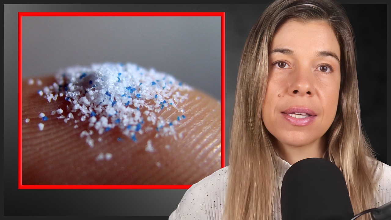
In this clip, Dr. Rhonda Patrick discusses the impact of microplastics and BPA on brain health, neurodevelopment, and neurodegenerative disease risk.

In this clip, Dr. Rhonda Patrick discusses magnesium supplementation, including dosing strategies and different forms' effectiveness and unique benefits.

In this clip, Dr. Martin Gibala delves into the cognitive advantages of engaging in high-intensity exercise.
-
In this clip, Dr. Rhonda Patrick discusses the impact of microplastics and BPA on brain health, neurodevelopment, and neurodegenerative disease risk.
-
In this clip, Dr. Rhonda Patrick discusses magnesium supplementation, including dosing strategies and different forms' effectiveness and unique benefits.
-
In this clip, Dr. Martin Gibala delves into the cognitive advantages of engaging in high-intensity exercise.
-
The blood-brain barrier is a collection of membranes that separate the central nervous system from the circulatory system. It permits the passage of nutrient...
-
In this clip, Dr. Axel Montagne describes the dire consequences of a dysfunctional blood-brain barrier.
-
In this clip, Dr. Axel Montagne highlights the blood-brain barrier changes that potentially contribute to Alzheimer's disease.
-
Dr. Axel Montagne discusses the importance of blood-brain barrier integrity, especially for people with APOE4, who have higher Alzheimer's disease risk.
-
Dr. Axel Montagne discusses blood-brain barrier dysfunction in dementia and early-stage age-related cognitive decline.
-
Rhonda Vitamin D Exercise Brain Alzheimer's Parkinson's Cancer Microbiome Cholesterol Omega-3 Skin Sulforaphane Protein NAD+ Moringa Blood-Brain Barrier CocoaDr. Rhonda Patrick answers audience questions on various health, nutrition, and science topics in this Q&A session.
-
Intestinal Permeability: the Bacterial link to Aging, Brain Barrier Dysfunction & Metabolic DisorderIntestinal Permeability Alzheimer's Gut Heart Disease Inflammation Depression Blood-Brain Barrier LipopolysaccharideDr. Patrick's keynote: compromised intestinal barrier affects human health—cardiometabolic function, neurological health, behavior, and more.
Topic Pages
-
Blood-brain barrier
The blood–brain barrier comprises tight-junctioned endothelial cells that mediate highly selective transcellular and paracellular exchange between blood and CNS.
-
Choline
Choline crosses the blood-brain barrier via a saturable, carrier-mediated low-affinity transporter to support acetylcholine synthesis.
-
Exercise and Cognitive Function
Exercise fortifies BBB tight-junctions, mitigates neuroinflammation, and consequently enhances synaptic plasticity, neurogenesis, and cognition.
-
Small vessel disease
Cerebral small-vessel disease mechanistically involves blood–brain barrier dysfunction that incites capillary leakage, neuroinflammation, and white-matter ischemia.
News & Publications
-
Exercise affects multiple physiological processes regulated by hormones, including stress responses and energy balance. Yet scientists still do not fully understand how some hormone precursors travel through the bloodstream during these demanding moments. A new study explored how vigorous exercise changes the way a major hormone precursor, called proopiomelanocortin (POMC), travels through the bloodstream and helps it reach sensitive tissues like the brain.
The study involved 15 healthy, endurance-trained adults who completed a 50-minute treadmill run at high intensity. The researchers focused on whether exercise encourages POMC to attach to small extracellular vesicles (often abbreviated to sEVs). These are microscopic, membrane-wrapped particles released by cells that can carry biological signals between tissues. They also tested the effect of this attachment on how easily POMC crosses cellular barriers.
- Most circulating POMC remained intact rather than being processed into smaller hormones such as ACTH or β-endorphin.
- Total POMC levels in blood also did not rise with exercise, nor did the number or size of sEVs. However, about four times more POMC was found attached to sEVs, and this shift partly reversed during recovery.
- Lab tests showed that making blood slightly more acidic, at levels similar to those seen during hard exercise, increased how much POMC attached to sEVs.
- POMC bound to sEVs crossed layers of endothelial cells (cells that line blood vessels) and a laboratory model of the blood–brain barrier more efficiently than free POMC.
- After crossing barriers in lab models, vesicle-bound POMC produced a measurable response in test cells.
To explain these results, the researchers turned to molecular modeling. Their analyses suggest that lower blood pH (higher acidity) causes a subtle shape change in POMC, exposing a region that binds more easily to receptors embedded in vesicle membranes. In other words, exercise-related acidity appears to make POMC better suited to latch onto sEVs, which then act as transport vehicles.
The study was small and focused on fit adults, and many experiments used laboratory models rather than living tissues. Still, the findings point to a previously unrecognized way exercise may fine-tune hormonal signaling. If confirmed, this vesicle-based transport system could help explain how physical stress rapidly reshapes communication between blood, immune cells, and the brain. In this clip, I discuss how vigorous exercise improves heart and brain health, enhances focus, and provides anti-cancer benefits.
-
Tiny brain hemorrhages found in 17% of people who've had a concussion—increasing their risk for dementia. jamanetwork.com
“Seeing stars,” “getting your bell rung,” and “knocked for a loop” are everyday phrases often used to describe experiencing a concussion. However, these expressions can downplay that even a “mild” concussion is a traumatic brain injury with the potential for lasting, harmful effects, including dementia. A recent study found that 17% of people who have had a concussion have microbleeds—tiny hemorrhages—in their brains.
The study involved more than 600 middle-aged adults with normal cognitive function. Researchers asked the participants if they had ever experienced a concussion—defined as a blow to the head where they lost consciousness—then assessed their brain health using magnetic resonance imaging (MRI). Then, they gave the participants various cognitive tests and asked about their sleep, mood, and overall health.
They found that more than one-third of the participants had experienced a concussion. Of these, 17% had evidence of brain microbleeds, and the more concussions they experienced, the more microbleeds they had. Those who had experienced a concussion tended to have poorer sleep and more gait disturbances, depression symptoms, and memory deficits than those without a concussion.
Microbleeds are markers of cerebral small vessel disease—age-related blood vessel impairments in the brain. These impairments compromise the blood-brain barrier, contributing to the development of cardiovascular disease, dementia, and stroke.
These findings suggest that even mild concussions damage the brain, increasing a person’s risk of dementia. Other studies have demonstrated that mild concussions cause acute and chronic brain damage, and people who experience three or more concussions tend to exhibit cognitive deficits that worsen with each subsequent concussion.
We’ve covered small vessel disease in great detail: - Listen to researcher Dr. Axel Montagne describe the prevalence of this condition as a cause of dementia. - Read more about the condition in our overview article.
-
Omega-3 fatty acids preserve blood-brain barrier integrity, maintaining cognitive function. onlinelibrary.wiley.com
The blood-brain barrier is a collection of membranes that separate the central nervous system from the peripheral circulation, facilitating the passage of nutrients and signaling compounds while excluding harmful substances. Loss of blood-brain barrier integrity increases the risk of many neurodegenerative diseases, including dementia and Alzheimer’s. A 2021 study found that having a higher omega-3 index preserves the blood-brain barrier and maintains cognitive function in older adults.
Researchers assessed the cognitive performance of 45 healthy older adults. They measured the participants' red blood cell omega-3 concentrations (“omega-3 index”) and evaluated their blood-brain barrier integrity using magnetic resonance imaging.
They found that participants with a higher omega-3 index had greater blood-brain barrier integrity than those with a lower index. They also performed better on tests of memory and language (functions most affected by Alzheimer’s disease) but not on executive function, speed, or motor control assessments.
These findings suggest that higher blood concentrations of omega-3 fatty acids (measured via the omega-3 index) preserve blood-brain barrier integrity, potentially protecting against cognitive losses. The omega-3 index measures the amount of the omega-3 fatty acids EPA and DHA as a percentage of total fatty acids in red blood cell membranes. Evidence suggests it is a reliable biomarker of sudden cardiac death risk and may provide a means of standardizing methodologies used in clinical trials, facilitating more accurate interpretation of clinical trial outcomes. Learn more about the omega-3 index in our overview article.
-
Aging undermines the brain's capacity for maintaining working memory, with subtle declines in neuron activity and connectivity in the prefrontal cortex. www.sciencedaily.com
As the brain ages, cognitive functions decline, with working memory – a form of short-term memory necessary for reasoning, decision-making, and behavior – among the earliest affected. The brain’s prefrontal cortex is crucial in working memory, showing sustained activity during memory-guided behavior. A recent study in mice found that aging reduces the prefrontal cortex’s ability to maintain working memory, with older mice showing less effective communication between memory-coding neurons.
Using imaging techniques to see how neurons behave during memory tasks involving touch and sound, researchers tracked brain activity in mice of varying ages. They also used light-based interventions to temporarily disrupt brain activity, observing how this affected the animals' ability to perform the tasks.
They discovered that with aging, the prefrontal cortex had fewer neurons responsible for managing actions, and the signals from these neurons weakened. Furthermore, whereas young mice used both broad and specific memory strategies, older mice mainly relied on specific strategies, indicating a subtle decline in the brain’s memory circuits over time.
The researchers also observed a drop in the resting brain connections among neurons managing actions, beginning around middle age. This drop in connections became even more pronounced when the mice were doing tasks, suggesting that the brain’s ability to retain thoughts might weaken with age. Additionally, the brains of middle-aged mice were more easily disrupted by light-based interventions, hinting at a greater risk to their thought-retention processes as they age.
The findings in this mouse study suggest that aging leads to a marked decrease in brain connectivity and neuron activity in the prefrontal cortex, affecting memory retention and making it more susceptible to disruptions. The blood-brain barrier is a critical player in brain connectivity, and its impairment during aging contributes to cognitive dysfunction. Learn more in this episode featuring Dr. Axel Montagne.
-
Pericytes are pivotal in the formation and storage of long-term memories. www.sciencedaily.com
Tiny contractile cells surrounding the brain’s capillaries called pericytes regulate vascular blood flow and maintain blood-brain barrier integrity. Pericytes detach from the blood vessels in aging, driving the pathophysiology of neurological dysfunction, vascular dementia, and stroke. A recent study in rodents shows that pericytes also play roles in long-term memory formation.
Researchers measured the amount of insulin-like growth factor 2 (IGF2) produced by various cells in the hippocampus of rodents. IGF2, a peptide hormone produced in multiple tissues, regulates growth during fetal development and participates in the cell cycle throughout the lifespan. After determining that pericytes contributed the greatest amount of hippocampal IGF2, they assessed learning’s influence on IGF2.
They found that learning increased pericyte IGF2 production in the hippocampus, especially in the dentate gyrus, a highly vascularized area responsible for episodic memory – long-term memory that involves conscious recollection of previous experiences and their associated contexts, such as sounds and smells. Animals lacking the ability to produce IGF2 in their pericytes exhibited poor learning and memory.
The detachment and loss of pericytes play a crucial role in the progression of cerebral small vessel disease and neurodegenerative disorders that involve blood-brain barrier dysfunction. These specialized endothelial cells envelop a significant portion, up to 80 percent, of the brain capillary surface area in the cortex and hippocampus of the human brain. They also enwrap the tiniest vessels constituting the blood-brain barrier.
Exercise mitigates the proinflammatory state that drives pericyte loss in aging, possibly providing a mechanism for exercise’s memory-enhancing effects. Learn more about links between exercise, pericytes, and brain health in this episode featuring Dr. Axel Montagne.
-
Fungi drive many chronic health conditions, including asthma, skin problems, gut disorders, and others. Recent research has highlighted potential links between the fungus Candida albicans and Alzheimer’s disease. A recent study in mice found that C. albicans triggers a dual-pronged defense system involving both the brain and the immune system; however, when elements of this system fail, the fungi can impair the blood-brain barrier and drive amyloid plaque formation.
Researchers injected mice with different strains of C. albicans and measured fungal levels in the animals' brains. They assessed their immune responses and examined their transendothelial electrical resistance – a crucial measure of the integrity and functionality of the endothelial layer, which forms the blood-brain barrier.
They found that Candida albicans employed enzymes called aspartic proteinases (Saps) to compromise blood-brain barrier integrity, facilitating fungal entry into the brain. Interestingly, the presence of Saps promoted the breakdown of amyloid precursor protein (APP) into smaller, potentially protective components to combat the fungal invasion.
The fungi also released candidalysin, a toxin that contributes to Candida albicans virulence. When microglia (brain immune cells) recognized the altered APP components and candidalysin, they synergized their efforts, driving the eradication of the fungus from the brain. However, if the microglia failed to recognize the APP or candidalysin, the fungus persisted in the brain, eliciting damage to neuronal tissues.
Amyloid precursor protein occurs naturally in the body, particularly in brain tissues. It participates in various cellular functions, including forming and maintaining synapses, which are essential for communication between nerve cells. Abnormal APP processing can promote amyloid-beta accumulation, a hallmark of Alzheimer’s disease.
These findings suggest the brain employs a complex defense system to protect itself against Candida albicans, a common fungus. However, the system’s failure may drive amyloid-beta accumulation and possibly Alzheimer’s disease. Learn more about amyloid-beta in this clip featuring Dr. Dale Bredesen.
-
Platelet-derived 'exerkines' produced during exercise promote neurogenesis, boosting brain health and cognition. www.sciencedaily.com
The benefits of physical activity on the brain’s aging process are widely known. Evidence suggests that exerkines, a class of molecules released into the bloodstream in response to exercise, drive many of these benefits. Findings from a recent study in mice found that PF4, an exerkine derived from platelets, promotes the production of hippocampal precursor cells in the brains of older mice.
Researchers injected PF4 into mice and assessed its effects on hippocampal neurogenesis (the growth of new brain cells). Then they investigated the effects of exercise on blood platelets.
They found that systemic elevation of PF4 levels mitigated age-related declines in brain regeneration and cognitive function, an effect that was dependent upon hippocampal neurogenesis. They also found that exercise triggered platelet activation, which in turn increased the production of hippocampal precursor cells in the brains of older mice.
These findings underscore the crucial role of platelets in mediating the rejuvenating effects of exercise on the aging brain. It also sheds light on the potential mechanisms that link physical activity with improved brain health in aging, with possible implications for people who are unable to exercise due to advanced age, mobility issues, or various health conditions.
Interestingly, heat stress also promotes PF4. In a study involving endurance cyclists exercising in hot conditions, PF4 increased by as much as 150 percent. Sauna use has similar effects on PF4. Learn about other effects of heat stress from sauna use in our overview article.
-
Microglia's Alzheimer's defense impaired by oral bacteria, study reveals. www.sciencedaily.com
Microglia, the brain’s resident immune cells, play a vital role in managing brain inflammation and neurodegenerative diseases by eliminating amyloid-beta, a harmful protein linked to Alzheimer’s disease, and forming barriers around insoluble amyloid-beta deposits. A new study in mice shows that oral bacteria over-stimulate microglia, impairing their anti-amyloid properties.
Researchers induced periodontal disease in mice by placing ligatures around their teeth, creating an environment conducive to bacterial growth. Then they examined the effects of the disease on the animals' gums, bones, and microglial cells. They found that the gum infections caused progressive periodontal disease and bone loss in the mice. In addition, the severity of periodontal disease correlated with increased microglial cell activation in the brain. Then, the researchers exposed microglial cells to bacteria from the animals' infected gums. They found that this exposure increased inflammation and changes in the cells' ability to interact with amyloid-beta.
These findings suggest that periodontal disease influences the brain’s immune response via changes in microglial activation and their interactions with amyloid-beta. This link between gum and brain health underscores the potential importance of oral hygiene in preventing or managing neuroinflammatory conditions like Alzheimer’s disease.
Related studies have found that oral bacteria colonize the brain and release toxins that disrupt the blood-brain barrier, impairing its function and increasing the risk of Alzheimer’s disease. Learn more about the role of blood-brain barrier dysfunction in Alzheimer’s disease in this episode featuring Dr. Axel Montagne.
-
Greater, more intense physical activity may preserve brain volume – especially in the hippocampus, an area of the brain involved in memory. Physichttps://neurosciencenews.com/physical-activity-neuroprotection-21177/al activity boosts brain health.
Being more physically active is associated with having larger brain volume, a 2022 study found. The greatest brain-enhancing effects were seen in those who engaged in moderate- to vigorous-intensity exercises.
The study involved more than 2,500 adults between the ages of 30 and 94 years. The participants wore wrist accelerometers to track their activity levels and underwent brain scans to assess their brain volume, cortical thickness, and gray matter density.
The scans revealed that physical activity had a marked, dose-dependent effect on the participants' brain health. Those with greater, more intense physical activity levels exhibited larger brain volumes – especially in the hippocampus, an area of the brain involved in memory – than those with lower, less intense levels. However, the beneficial effects of physical activity on brain health were particularly evident when comparing the brain volumes of those who engaged in moderate physical activity to those who were sedentary, suggesting that even small increases in physical activity can boost brain health and reduce brain volume losses associated with aging.
The beneficial effects of physical activity may be related to exercise-induced production of brain-derived neurotrophic factor (BDNF), a protein that controls and promotes the growth of new neurons. BDNF is active in the hippocampus, cortex, cerebellum, and basal forebrain – areas involved in learning, long term memory, and executive function. Learn more about the brain-boosting effects of BDNF in our comprehensive overview article.
-
High-dose omega-3 treatment expression of Mfd2a omega-3 DHA transporter in retina and blood vessels www.sciencedirect.com
From the publication:
Most importantly, our results revealed that high-dose fish oil supplementation was able to induce the expression of Mfsd2a, a major DHA transporter in the retina on both transcriptional and translational levels, simultaneously increasing the Mfsd2a expression on the blood vessels after only three weeks of treatment. It was suggested that the low expression of Mfsd2a in the retina might be one reason why DHA therapy fails to alleviate the symptoms of diabetic retinopathy (DR) and that the combined use of Mfsd2a overexpression and DHA therapy may have synergistic effects. It is thus conceivable that the high dose fish oil supplementation can serve as a potential adjuvant in the therapies or as a prophylactic in the early stages of disease for the impaired blood-retinal barrier through the up-regulation of Mfsd2a expression.
-
Just two hours of air pollution exposure interferes with regular brain activity. www.sciencedaily.com
Even short-term exposure to air pollution affects brain function, a new study shows. People exposed to diesel exhaust for just two hours had reduced connectivity in areas of the brain associated with attention and focus.
Researchers exposed 25 healthy adults to diesel exhaust and filtered air for two hours, with a two-week break between each exposure. Before and after the exposures, the researchers measured activity in a region of the participants' brains called the default mode network.
They found that compared to pre-exposure, participants had reduced connectivity throughout the default mode network of their brains after brief exposure to diesel exhaust. There were no changes in connectivity following exposure to filtered air.
The default mode network is a collection of interconnected neural structures involved in attention and focus. Disturbances in default mode network connectivity are associated with poor working memory, reduced performance, and work-related productivity losses.
Sulforaphane, a bioactive compound derived from broccoli – and particularly abundant in broccoli sprouts – promotes urinary excretion of some components of air pollution. Learn more in the clip featuring Dr. Jed Fahey.
-
Having kidney disease increases the risk of bleeding in the brain. www.ncbi.nlm.nih.gov
A 2020 study found that kidney disease increases the risk of having tiny brain hemorrhages called microbleeds. People with kidney disease were five times more likely to have microbleeds than those with normal kidney function.
Researchers studied the effects of kidney disease on cultured brain endothelial cells and in mice. They also tracked the progression of microbleeds in people who had kidney disease or were healthy.
They found that exposing the cells to urea – a byproduct of protein metabolism that builds up in the blood during kidney disease – damaged the endothelial cells' integrity, compromising their capacity to maintain the blood-brain barrier. The mice with kidney disease had twice as many microbleeds in their brains as healthy mice and showed signs of increased blood-brain barrier permeability. Fifty percent of the people who had kidney disease had microbleeds, whereas just 10 percent of those who were healthy had microbleeds.
Microbleeds are cerebral microhemorrhages that occur due to cerebral amyloid angiopathy, chronic hypertension, and other vascular conditions. Having a high microbleed count is associated with impaired cognitive function. Microbleeds are a hallmark of cerebral small vessel disease.
Evidence suggests that omega-3 fatty acids reduce the risk of chronic kidney disease. Omega-3 fatty acids may protect the kidneys by improving endothelial function, reducing blood pressure, and maintaining healthy blood lipids. Learn more about the benefits of omega-3s in our overview article.
-
Choline deficiency may increase the risk of Alzheimer's disease by promoting the formation of amyloid-beta and tau. www.sciencedaily.com
A new study in mice shows that choline deficiency increases the risk of Alzheimer’s disease. Researchers fed mice that are predisposed to Alzheimer’s disease either a choline-rich or choline-poor diet for seven months, starting in midlife until late life. They subjected the mice to motor and memory skills tests, and then they examined the animals' brains and other organs.
They found that mice that ate a choline-poor diet had higher brain levels of amyloid-beta and tau – two proteins implicated in the pathogenesis of Alzheimer’s disease – than those that ate a choline-rich diet. The mice that ate a choline-poor diet also gained weight, showed signs of altered metabolism, liver damage, and enlarged hearts, and performed poorly on motor skills tests.
Choline is an essential nutrient that supports the production of acetylcholine, a neurotransmitter involved in neurogenesis, synapse formation, learning, and memory. It is produced in the liver and is also found in foods such as eggs, meat, fish, beans, and nuts and as a dietary supplement. Most people living in the United States don’t consume enough choline – 550 milligrams per day for men and 425 milligrams per day for women – potentially increasing their risk for various diseases.
These findings suggest that choline deficiency increases the risk of Alzheimer’s disease and damages vital organs in mice. For a tasty way to get more choline into your diet, try this low-carb, choline-rich lemon tart.
-
Aerobic exercise reduces symptoms of anxiety. bmchealthservres.biomedcentral.com
Aerobic exercise – especially high-intensity exercise – reduces symptoms of anxiety, an analysis of 15 studies shows. These anti-anxiety effects endured for several months after cessation of the exercise.
Reviewers analyzed data from 15 studies that investigated the effects of low- or high-intensity exercise on anxiety symptoms. All the participants in the studies had some degree of anxiety, with their conditions falling on a spectrum that included anxiety disorders, raised anxiety levels, and raised anxiety sensitivity – a condition in which a person feels anxious about the physical symptoms that often accompany anxiety. People on waiting lists for anxiety treatment who did not exercise served as comparisons.
They found that participants who engaged in both low- and high-intensity aerobic exercise experienced greater improvements in their anxiety than non-exercising people on treatment waiting lists. High-intensity exercise reduced anxiety symptoms more effectively than low-intensity exercise. The various interventions lasted between 10 weeks and six months, with participants exercising three times a week, on average.
Multiple mechanisms may be responsible for the anti-anxiety effects of exercise. For example, high-intensity exercise promotes the production of lactate, a byproduct of glucose metabolism that participates in the production of neurotransmitters, such as norepinephrine and serotonin. Low norepinephrine and serotonin levels can drive anxiety and the inability to handle stressful situations. Learn more about lactate and its effects on the brain in this episode featuring Dr. George Brooks.
-
Greater visceral and total body fat associated with vascular brain injury and impaired cognitive scores www.sciencedaily.com
From the article:
In the study, 9,166 participants were measured by bioelectrical impedance analysis to assess their total body fat.
As well, 6,733 of the participants underwent magnetic resonance imaging (MRI) to measure abdominal fat packed around the organs known as visceral fat, and the MRI also assessed vascular brain injury – areas in the brain affected by reduced blood flow to the brain.
[…]
Co-author Eric Smith, a neurologist, scientist and an associate professor of clinical neurosciences at the University of Calgary, said that “preserving cognitive function is one of the best ways to prevent dementia in old age. This study suggests that one of the ways that good nutrition and physical activity prevent dementia may be by maintaining healthy weight and body fat percentage.”
-
Four pathologies of SVD associate with dementia: white matter hyperintensities, cerebral microbleeds, lacunar infarcts, enlarged perivascular spaces www.sciencedaily.com
The four pathologies that were detectable by MRI included:
- moderate-to-severe white matter hyperintensities - blood pressure is a risk factor for this one
- deep cerebral microbleeds
- lacunar infarcts
- enlarged perivascular space.
From the article:
The subjects were given cognitive tests and brain MRIs. The MRIs were examined for four main components of small vessel disease (SVD). These four components, which include evidence of microbleeds and minor strokes, then were added to create a total SVD score. The score ranges from zero points (no SVD) to 4 points (severe SVD).
The study found that that 61 percent of the subjects had zero points on the total SVD score, 20 percent had 1 point, 12 percent had 2 points, 5 percent had 3 points and 2 percent had 4 points. The higher the SVD score, the greater the cognitive decline. Researchers also found that each individual component of SVD predicted cognitive decline as well as the total SVD score did.
-
Small-vessel disease could provoke amyloid pathology while Alzheimer's-associated cerebral amyloid pathology may lead to auxiliary vascular damage www.sciencedaily.com
Cerebral small vessel disease is associated with amyloid-beta deposition in the brain, especially among APOE4 carriers.
Small vessel disease is a collection of conditions characterized by damage to arterioles and capillaries, resulting in reduced or interrupted blood flow to the affected organ. These conditions typically affect organs that receive substantial blood flow, such as the brain, kidney, and retina, and are principal drivers of chronic diseases such as strokes, renal failure, dementia, and blindness. Findings from a 2014 study suggest that people who have small vessel disease in the brain exhibit greater deposition of amyloid-beta plaques, especially if they are carriers of the APOE4 gene.
Amyloid-beta is a toxic 42-amino acid peptide that aggregates and forms plaques in the brain with age. Amyloid-beta deposition is associated with Alzheimer’s disease, a progressive neurodegenerative disease that can occur in middle or old age and is the most common cause of dementia.
APOE is a protein involved in lipid transport. A variant in the APOE gene, called apolipoprotein E4 (APOE4), is the major genetic risk factor for Alzheimer’s disease. Having one APOE4 allele increases a person’s Alzheimer’s disease risk as much as threefold; carrying two APOE4 alleles increases risk as much as 15-fold.
The cross-sectional study included more than 900 patients enrolled in the Amsterdam Dementia Cohort study who had been diagnosed as having Alzheimer’s disease, vascular dementia, or self-reported memory complaints. The investigators analyzed the patients' cerebrospinal fluid for the presence of amyloid-beta and other markers of Alzheimer’s disease and genotyped the patients to assess APOE status.
They also performed magnetic resonance imaging (MRI) of the patients' brains to identify the presence of white matter hyperintensities and microbleeds. White matter hyperintensities, areas in the brain that appear as intense white spots on MRIs, are often indicators of cerebral small vessel disease and are considered a risk factor for dementia. Microbleeds are small, chronic hemorrhages that are indicative of cerebral amyloid angiopathy, a condition in which amyloid-beta accumulates on the walls of brain arteries.
They found that patients with Alzheimer’s disease had lower levels of amyloid-beta in their cerebrospinal fluid, an effect that was more pronounced among APOE4 carriers. Patients with low amyloid-beta levels in their cerebrospinal fluid were more likely to have white matter hyperintensities and microbleeds, indicating a direct relationship between a pathological hallmark of Alzheimer’s disease and small vessel disease.
These findings suggest that Alzheimer’s disease and small vessel disease are intrinsically linked, especially among APOE4 carriers. [Learn more about small vessel disease in our overview article.](Coming soon)
-
Anesthesia and Surgery Impair Blood–Brain Barrier and Cognitive Function www.frontiersin.org
Surgery and anesthesia may contribute to cognitive impairment.
Anesthesia, a medical treatment that involves the use of anesthetic drugs, is a widely accepted strategy for managing pain during surgery. Evidence suggests that mice that have undergone surgery that involved anesthetic treatment exhibited signs of postoperative cognitive impairment. Findings from a 2017 study suggest that surgery in combination with the anesthetic drug isoflurane induces blood-brain barrier dysfunction, contributing to cognitive impairment.
Anesthetic drugs can be administered orally, intravenously, or via inhalation. Isoflurane is one of the most common inhaled anesthetics used in humans. Scientists don’t fully understand the mechanisms by which inhaled anesthetics work, but they appear to affect the central nervous system by depressing neurotransmission pathways. Previous research has shown that isoflurane increases several markers of inflammation.
The investigators wanted to determine whether the effects of surgery and anesthesia on blood-brain barrier permeability and cognitive function were related to age and/or the action of interleukin-6 (IL-6), a proinflammatory molecule. The experiment involved three types of mice: young female mice, old female mice, and young female mice that don’t carry the gene for IL-6. Previous research has shown that female mice are more vulnerable to the cognitive impairment associated with surgery and anesthesia. Half of the mice underwent a simple surgical procedure under anesthesia (isoflurane), while the remaining half did not. After the mice that had had surgery recovered, the investigators assessed all the animals' cognitive performance via a maze test, measured IL-6 in the animals' blood, and quantified proteins involved in maintaining blood-brain barrier integrity in the animals' brains.
They found that the mice that underwent anesthesia and surgery had greater blood-brain permeability and higher IL-6 levels than those that did not undergo the procedures. Notably, permeability increased as IL-6 levels and age increased. Levels of proteins involved in maintaining blood-brain barrier integrity were lower in the mice that underwent the procedure, and older mice were more likely to experience cognitive deficits after the procedures than younger mice.
These findings suggest that surgery with anesthesia induces cognitive decline in an age-dependent manner, and this decline may be due to inflammatory processes that drive blood-brain barrier dysfunction. Learn more about the blood-brain barrier in our new overview article.
-
Repeated head injury damage the blood-brain barrier and produce an autoimmune response that promotes neurological disease www.sciencedaily.com
From the article:
A new study suggests that brain injury from repeat blows to the head – observed among football players and soldiers – might not be a traumatic phenomenon, but an autoimmune phenomenon. It indicates that brain injury may be the result of an out-of-control immune response, much like multiple sclerosis. This is an entirely new way of thinking about how trauma could cause long term degeneration and opens the door to investigating a vaccine/drug to prevent head trauma.
[…]
Researchers found that S100B, a well-accepted protein biomarker for traumatic brain injury, was present in varying degrees in the blood samples of the 67 football players after every game – even though none of them suffered a concussion. This demonstrates that even the most routine hits have some impact on the blood-brain barrier and possibly the brain itself, Bazarian said.
-
Blood-brain barrier may be impaired by even mild head trauma: 50% of rugby players and a similar amount in MMA fighters show damage after a season Blood-Brain Barrier Damage Occurs Even With Mild Head Trauma
Mild head trauma not resulting in a concussion may cause damage to the blood-brain barrier.
From the article:
In the study, which was published online Sept. 5 in the Journal of Neurotrauma, scientists scanned the brains of adolescent and adult rugby players with a special type of magnetic resonance imaging. They found damage to the protective barrier that separates the brain from bloodborne pathogens and toxins in roughly half of adolescent rugby players after a full season — even those who did not report a concussion. Professional mixed martial arts fighters showed similar damage after a fight.
Extremely frequent BBB disruption in rugby players:
Ten of 19 adolescent rugby players showed signs of blood-brain barrier disruption by the end of the season. The barrier breakdown appeared on the scans as red blips concentrated throughout the inner regions of the brain. The researchers also scanned eight college rugby players after a match and saw disruptions of the barrier in two of them. Notably, the injuries most study participants experienced were below the current bar for mild head trauma since they did not suffer a concussion.
-
Effective clinical treatment of depression may require addressing underlying loss of blood-brain barrier integrity www.sciencedaily.com
From the article:
The researchers examined blood-brain barrier cells in depressed stressed mice, resilient stressed mice, and control mice. Their observations show that the epigenetic processes that allow the expression of the claudin-5 gene are more readily activated in resilient mice. They also observed that the resilient mice produce less of one of the proteins that inhibit expression of the claudin-5 gene.
Conversely, depressed stressed mice express more of an enzyme called HDAC1 that triggers a loss of claudin-5. “When a chemical compound is used to block HDAC1, the depressive mice produce more claudin-5 and their social interactions spontaneously increase,” says Professor Ménard.
-
Restoring integrity of blood-brain barrier critical for epilepsy: blood-brain barrier disruption was a key driver of seizure activity www.sciencedaily.com
Probably one of the better known and highly effective non-pharmacological approaches to changing seizure threshold is achieving nutritional ketosis via a ketogenic diet. Which begs the question… what effect does nutritional ketosis have on maintenance of the blood-brain barrier?
From the article:
Scientists have announced a significant advance in our understanding of epilepsy, as they have identified a potential method of preventing damaging seizure activity. Brain cells are nourished by an intricate network of capillaries that forms the so-called blood-brain barrier (BBB). Fundamentally, it is disruption to the integrity of these capillaries and the BBB that a group of scientists believe is a key driver of seizure activity in humans. Promisingly though, their new research shows that restoring that integrity can prevent seizures.
Animal and human evidence:
Importantly, the work was translational in nature and included both basic and clinical research arms involving patients diagnosed with epilepsy. Using similar techniques in humans and in pre-clinical models, the scientists were able to show that BBB disruption was a key driver of seizure activity.
-
Air pollution may promote the production of autoantibodies against tight-junctions and neural proteins: neuroinflammation in children in Mexico City www.sciencedaily.com
Exposure to air pollution promotes the production of autoantibodies against tight-junctions of the blood-brain barrier.
Separately, evidence has also shown that even very young children show evidence of amyloid-beta build up under these conditions.
From the article:
The study found when air particulate matter and their components such as metals are inhaled or swallowed, they pass through damaged barriers, including respiratory, gastrointestinal and the blood-brain barriers and can result in long-lasting harmful effects.
The results found that the children living in Mexico City had significantly higher serum and cerebrospinal fluid levels of autoantibodies against key tight-junction and neural proteins, as well as combustion-related metals.
“We asked why a clinically healthy kid is making autoantibodies against their own brain components,” Calderón-Garcidueñas said. “That is indicative of damage to barriers that keep antigens and neurotoxins away from the brain. Brain autoantibodies are one of the features in the brains of people who have neuroinflammatory diseases like multiple sclerosis.”
-
Routine tests ignore breakdown of blood-brain barrier and altered cerebral blood flow that precede Alzheimer's diagnosis www.sciencedaily.com
From the article:
“Cognitive impairment, and accumulation in the brain of the abnormal proteins amyloid and tau, are what we currently rely upon to diagnose Alzheimer’s disease, but blood-brain barrier breakdown and cerebral blood flow changes can be seen much earlier,” said Berislav Zlokovic, the Mary Hayley and Selim Zilkha Chair in Alzheimer’s Disease Research at the Keck School of Medicine of USC. “This shows why healthy blood vessels are so important for normal brain functioning.”
[…]
BBB leaks can be detected with an intravenously administered contrast substance in concert with magnetic resonance imaging. Brain microbleeds, another sign of leakage, also can be picked up with MRI. A slowdown in the brain’s uptake of glucose, visible via PET scan, can be a another result of BBB breakdown. Zlokovic notes that these aren’t tests routinely offered at a doctor’s office.
-
Model of blood-brain barrier malfunctions when stem cells are derived from cells of patients with Huntington's disease www.sciencedaily.com
From the article:
For their study, a team led by Cedars-Sinai investigators generated stem cells known as induced pluripotent stem cells, which can produce any type of cell, using an individual adult’s blood samples. They used these special cells to make neurons, blood-vessel linings and support cells that together make up the blood-brain barrier. The team then placed the various types of cells inside Organ-Chips, which recreated the body’s microenvironment with the natural physiology and mechanical forces that cells experience within the human body.
The living cells soon formed a functioning unit of a blood-brain barrier that functions as it does in the body, including blocking entry of certain drugs. Significantly, when this blood-brain barrier was derived from cells of patients with Huntington’s disease or Allan-Herndon-Dudley syndrome, a rare congenital neurological disorder, the barrier malfunctioned in the same way that it does in patients with these diseases.
-
Inherited Parkinson's LRRK2 mutations promote excessive inflammatory cytokines that cross blood-brain barrier, trigger neuron damage via microglia www.sciencedaily.com
From the article:
Suspecting that the LRRK2 mutations might be acting outside of the brain, the researchers used an agent – the outer shell of bacteria, called lippopolysaccharide (LPS) – that causes an immune reaction. LPS itself does not pass into the brain, nor do the immune cells it activates, which made it ideal for testing whether this second hit was acting directly in the brain.
When the researchers gave the bacterial fragments to the mice carrying the two most common LRRK2 gene mutations, the immune reaction became a “cytokine storm,” with inflammatory mediators rising to levels that 3-5 times higher than a normal reaction to LPS. These inflammatory mediators were produced by T and B immune cells expressing the LRRK2 mutation.
Despite the fact that LPS did not cross the blood-brain barrier, the researchers showed that the elevated cytokines were able to enter the brain, creating an environment that caused the microglia to activate pathologically and destroy the brain region involved in movement.
-
Repurposed chemo drug acting on the blood-brain barrier shows dose-dependent increase of dopamine in Parkinson's disease patients www.sciencedaily.com
Repurposed chemo drug inactivates protein that destroy’s the blood-brain barrier in Parkinson’s disease:
The current part of the study just published, examined the cerebrospinal fluid of patients via epigenomics, which is a systematic analysis of the global state of gene expression, in correlation with continuing clinical outcomes. The new analysis helps explain the clinical findings.
Nilotinib inactivated a protein (DDR1) that was destroying the ability of the blood brain barrier to function properly. When DDR1 was inhibited, normal transport of molecules in and out of the brain filter resumed, and inflammation declined to the point that dopamine, the neurotransmitter depleted by the disease process, was being produced again.
After 27 months, nilotinib was found to be safe, and patients who received nilotinib showed a dose-dependent increase of dopamine, the chemical lost as a result of neuronal destruction.
First study to show blood-brain barrier as therapeutic target for Parkinson’s disease:
“Not only does nilotinib flip on the brain’s garbage disposal system to eliminate bad toxic proteins, but it appears to also repair the blood brain barrier to allow this toxic waste to leave the brain and to allow nutrients in,” Moussa explains. “Parkinson’s disease is generally believed to involve mitochondrial or energy deficits that can be caused by environmental toxins or by toxic protein accumulation; it has never been identified as a vascular disease.”
“To our knowledge, this is the first study to show that the body’s blood brain barrier potentially offers a target for the treatment for Parkinson’s disease,” Moussa says.
-
Amyloid-beta can travel, cancer-like, to brain from other parts of body: insights from parabiosis experiments www.sciencedaily.com
From the article:
The scientists demonstrated this cancer-like mobility through a technique called parabiosis: surgically attaching two specimens together so they share the same blood supply for several months. […] Normal mice that had been joined to genetically modified partners for a year “contracted” Alzheimer’s disease. Song says the amyloid-beta traveled from the genetically-modified mice to the brains of their normal partners, where it accumulated and began to inflict damage.
The problem is partly due to an increased permissiveness of the blood-brain barrier as we age that allows entry from other parts of the body:
“The blood-brain barrier weakens as we age,” says Song, a Canada Research Chair in Alzheimer’s Disease and the Jack Brown and Family Professor. “That might allow more amyloid beta to infiltrate the brain, supplementing what is produced by the brain itself and accelerating the deterioration.”
-
Microbiota play important role in preventing blood-brain barrier permeability: germ-free mice have greater leakiness, resolved with fecal transplant www.sciencedaily.com
From the article:
The investigators reached this conclusion by comparing the integrity and development of the blood-brain barrier between two groups of mice: the first group was raised in an environment where they were exposed to normal bacteria, and the second (called germ-free mice) was kept in a sterile environment without any bacteria.
“We showed that the presence of the maternal gut microbiota during late pregnancy blocked the passage of labeled antibodies from the circulation into the brain parenchyma of the growing fetus,” says first author Dr. Viorica Braniste at the Department of Microbiology, Tumor and Cell Biology at Karolinska Institutet. “In contrast, in age-matched fetuses from germ-free mothers, these labeled antibodies easily crossed the blood-brain barrier and was detected within the brain parenchyma.”
However, it can be resolved with fecal transplant:
Interestingly, this ‘leakiness’ could be abrogated if the mice were exposed to fecal transplantation of normal gut microbes.
-
Chronic systemic inflammation induced by lipopolysaccharide can cause microglia to attack the blood-brain barrier www.sciencedaily.com
Obesity promotes circulation of lipopolysaccharide. In animals, chronic systemic inflammation, experimentally induced by injection with LPS, also known as “LPS challenge,” can cause microglia into the brain to switch from protecting the blood-brain barrier to damaging it.
From the article:
Nearly 50 percent of all dementias, including Alzheimer’s, begins with the breakdown of the smallest blood vessels in the brain and their protective “gatekeeper cells,” according to a Keck School of Medicine of USC study.
[…]
A key point of interest was the systemic inflammation induced by injecting the mice with an inflammation-inducing substance. Such injections resulted in the movement of microglia to the blood vessels and increased the permeability of the blood-brain barrier within a few days. Then, the microglia initially acted to protect the blood-brain barrier and limit increases in permeability, but as inflammation progressed, the microglia reversed their behavior by attacking the components of the blood-brain barrier, thus increasing the barrier’s permeability. The subsequent leakage of molecules into the brain had the potential to cause widespread inflammation in the brain and consequent damage to neurons (cells of the nerves).
-
50% of all dementias and Alzheimers may start with leaky blood-brain barrier www.sciencedaily.com
From the article:
“Many scientists have focused their Alzheimer’s disease research on the buildup of toxic amyloid and tau proteins in the brain, but this study and others from my lab show that the problem starts earlier – with leaky blood vessels in the brain,” said Berislav Zlokovic, senior author of the study
Reducing fibrinogen that enters the brain through leaky gatekeeping may be important for preventing decline:
Fibrinogen develops blood clots so wounds can heal. When gatekeeper cells are compromised, an unhealthy amount of fibrinogen slinks into the brain and causes white matter and brain structures, including axons (nerve fibers) and oligodendrocytes (cells that produces myelin), to die.
-
Spike proteins that extrude from SARS-CoV-2 promote inflammatory responses on the endothelial cells that form the blood-brain barrier. www.sciencedaily.com
SARS-CoV-2, the virus that causes COVID-19, disrupts the blood-brain barrier.
COVID-19 is widely regarded as a respiratory illness, but evidence suggests it affects multiple organ systems, including the central nervous system. For example, some people with COVID-19 experience headaches, nausea, vomiting, or “brain fog” – indicators of neurological involvement. Evidence from a 2020 study suggests that SARS-CoV-2, the virus that causes COVID-19, disrupts the blood-brain barrier.
SARS-CoV-2 enters cells via the angiotensin-converting enzyme 2 (ACE2), a protein that is widespread among the body’s tissues and plays important roles in blood pressure control. Once inside the cell, SARS-CoV-2 replicates, triggering a robust immune response and eliciting widespread inflammation.
The blood-brain barrier, a semi-permeable barrier that separates the blood from the brain’s extracellular fluid, prevents the entry of neurotoxic substances into the brain. Disruption of the blood-brain barrier has been implicated in the pathogenesis of neurodegenerative diseases, including Alzheimer’s disease, Parkinson’s disease, amyotrophic lateral sclerosis, and multiple sclerosis, among others.
The investigators first examined postmortem brain tissue from healthy people as well as people who had been diagnosed with hypertension (high blood pressure) or dementia to identify the presence of ACE2 in the brain blood vessels. They found that not only was ACE2 present in the blood vessels, but it was particularly abundant in people with hypertension or dementia. Then, using an in vitro model of the blood-brain barrier, they assessed the effects of exposure to the SARS-CoV-2 spike protein, the primary infectious particle on the virus. They found that exposure to the spike protein impaired blood-brain barrier function and integrity. Finally, using a tissue model that mimics the movement of fluid in the barrier, they found that the spike protein increased barrier permeability.
These findings suggest that SARS-CoV-2 binds to ACE2 receptors in the brain and impairs blood-brain barrier function and integrity. These effects may be exacerbated in people with co-existing illnesses such as hypertension or dementia.
-
Leaky blood-brain barrier caused by cerebral small vessel disease increases the prevalence of white-matter hyperintensities and brain damage www.sciencedaily.com
Poor blood-brain barrier integrity drives white matter losses.
White matter hyperintensities are areas in the brain that appear as intense white spots on magnetic resonance imaging (MRI) scans. They are often indicators of cerebral small blood vessel disease and are considered a risk factor for dementia. A 2021 study found that breaches in blood-brain barrier integrity are associated with brain tissue losses and precede the appearance of white matter hyperintensities.
The blood-brain barrier, a specialized system of endothelial cells that shields the brain from toxins present in the blood, supplies the brain’s tissues with vital nutrients and substances necessary for neuronal and metabolic function. The structural integrity of the blood-brain barrier is therefore critical for homeostatic maintenance of the brain microenvironment.
The study involved 43 patients (average age 58 years) who had been diagnosed with cerebral small vessel disease, as evidenced by having experienced a stroke or demonstrating mild cognitive impairment. At the beginning of the study and two years later, participants underwent a variety of MRI techniques that quantified their overall blood-brain barrier permeability as well as the areas surrounding white matter hyperintensities.
The MRIs revealed that participants who had the greatest amount of leaky brain tissue at the beginning of the study exhibited greater white matter tissue losses two years later. These tissue losses translated to greater permeability, a phenomenon particularly evident in the areas surrounding the brain lesions associated with white matter hyperintensities.
These findings suggest that losses in blood-brain barrier integrity damage brain tissue, driving increased permeability and white matter losses. In turn, these changes potentiate the disease processes associated with cerebral small vessel disease. Learn more about the blood-brain barrier in our overview article.
-
People with more severe leakage of blood-brain barrier have worse memory problems, potentially a separate or very early process in Alzheimer's disease www.sciencedaily.com
From the article:
USC’s five-year study, which involved 161 older adults, showed that people with the worst memory problems also had the most leakage in their brain’s blood vessels – regardless of whether abnormal proteins amyloid and tau were present.
“The fact that we’re seeing the blood vessels leaking, independent of tau and independent of amyloid, when people have cognitive impairment on a mild level, suggests it could be a totally separate process or a very early process,” said senior author Berislav Zlokovic, director of the Zilkha Neurogenetic Institute at the Keck School of Medicine of USC. “That was surprising that this blood-brain barrier breakdown is occurring independently.”
-
Alzheimer’s risk allele APOE4 triggers early breakdowns in blood-brain barrier, predicting cognitive decline www.sciencedaily.com
From the article:
Severe damage to vascular cells called pericytes was linked to more severe cognitive problems in APOE4 carriers. APOE4 seems to speed up breakdown of the blood-brain barrier by activating an inflammatory pathway in blood vessels, which is associated with pericyte injury."
[…]
Zlokovic’s previous research shows that people who develop early memory problems also experience the most leakage in their brain’s blood vessels – independent of amyloid plaque or tau, two common contributors to Alzheimer’s. The leakage starts when cells called pericytes, which line the walls of blood vessels in the brain and maintain blood-brain barrier integrity, are damaged.
[…]
In participants who had the APOE4 gene, researchers found damaged capillaries in the brain’s memory center, the hippocampus and medial temporal lobe. The damage correlated with increased levels of a protein that causes inflammation, cyclophilin A – an early sign of the disease in people already at higher risk of developing Alzheimer’s.
-
Inhibiting the Mfsd2a transporter that brings omega-3 DHA into the brain promotes leakage of brain barrier www.sciencedaily.com
Impaired transport of DHA disrupts the blood-brain barrier.
Lipid rafts – cholesterol-filled “bubbles” found in the cell membrane – serve as staging areas for many cellular activities. One type of lipid raft, called caveolae, facilitates the transport of substances across the membrane of endothelial cells. Findings from a 2017 study demonstrate that suppression of caveolae-mediated transport in brain endothelial cells protects the integrity of the blood-brain barrier.
The blood-brain barrier is a highly selective semi-permeable barrier made up of endothelial cells connected via tight junctions. This barrier separates the circulating blood from the brain’s extracellular fluid and prevents the entry of substances that may be neurotoxic. Disruption of the blood-brain barrier has been implicated in the pathogenesis of neurodegenerative diseases, including Alzheimer’s disease, Parkinson’s disease, amyotrophic lateral sclerosis, and multiple sclerosis, among others.
The investigators' previous research showed that a critical player in blood-brain barrier function is Mfsd2a, a transmembrane protein found exclusively on the endothelial cells that line blood vessels on the barrier. Mfsd2a participates in lipid transport and is the sole means by which lysophospholipid DHA, the brain’s preferred form of DHA (a type of omega-3 fatty acid) is delivered to the brain.
Using mice that carried a mutation that blocked Mfsd2a’s capacity to transport DHA, the investigators assessed blood-brain barrier function as well as caveolae formation and activity in the animals' brains. Then they compared the lipid composition of brain endothelial cells to lung epithelial cells, which lack Mfsd2a.
They found that mice that lacked Mfsd2a function had leakier blood-brain barriers and greater caveolae formation and activity than normal mice. They also found that brain endothelial cells had higher lipid concentrations than lung epithelial cells. The most abundant lipid in the brain endothelial cells was DHA, which was found in concentrations that were two to five times higher.
These findings suggest that Mfsd2a-mediated transport of lipids, particularly DHA, impairs caveolae activity, thereby preserving blood-brain integrity. Learn more about links between Mfsd2a, DHA, and brain health in this open-access peer-reviewed article by Dr. Rhonda Patrick..
[Learn more about the blood-brain barrier in our overview article.](LINK)
-
Reduced expression of genes involved in integrity of blood-brain barrier, intestinal barrier in autism: 75% have reduced barrier-forming components www.sciencedaily.com
From the article:
The group analyzed postmortem cerebral cortex and cerebellum tissues from 33 individuals – 8 with ASD, 10 with schizophrenia and 15 healthy controls. Altered expression of genes associated with blood-brain-barrier integrity and function and with inflammation was detected in ASD tissue samples, supporting the hypothesis that an impaired blood-brain barrier associated with neuroinflammation contributes to ASD.
In keeping with the hypothesis that the interplay within the gut-brain axis is a crucial component in the development of neurodevelopmental disorders, the group also analyzed intestinal epithelial tissue from 12 individuals with ASD and 9 without such disorders. That analysis revealed that 75 percent of the individuals affected by ASD had reduced expression of barrier-forming cellular components, compared with controls, and 66 percent showed a higher expression of molecules that increase intestinal permeability.
-
Obesity can break down our protective blood brain barrier resulting in problems with learning and memory: overactivity of adenosine in obesity www.sciencedaily.comBrain Alzheimer's Obesity Diabetes Coffee Insulin Blood Sugar Blood-Brain Barrier Lipopolysaccharide
Diet-induced insulin resistance caused blood vessels to become leaky which impaired blood and oxygen flow to a brain region involved in learning and memory (animal evidence).
Obesity and insulin resistance are associated with a leaky blood-brain barrier. This new animal study found that a high-sugar combined with a high-fat diet caused shrinkage of the tight junctions between endothelial cells that make up the blood-brain barrier and actual holes in those cells.
Obesity is known to increase toll-like receptor activation through a variety of mechanisms. One of the mechanisms is through through associated increases of circulating of lipopolysaccharide. Another mechanism is the leaking of fatty acids from fatty acids, triggering toll-like receptors through the recognition of damage-associated molecular patterns (DAMPs). (See “obesity” section of toll-like receptor article.)
Furthermore, LPS challenge in animal studies induces microglia in the brain to attack and damage the blood-brain barrier.
Blocking adenosine may play a role in preventing impairment of the blood-brain barrier by diet-induced obesityHowever, adenosine, which helps us sleep and helps regulate our blood pressure and is blocked by caffeine may play a role in preventing some of the damaging effects obesity has on the blood-brain barrier, which promotes dementia.
From the article:
They knew that chronic activation of the receptor Adora2a [an adenosine receptor] on the endothelial cells that line this important barrier in our brain can let factors from the blood enter the brain and affect the function of our neurons.
Now Medical College of Georgia scientists have shown that when they block Adora2a in a model of diet-induced obesity, this important barrier function is maintained.
[…]
In the brain, adenosine is a neurotransmitter that helps us sleep and helps regulate our blood pressure; in the body it’s also a component of the cell fuel adenosine triphosphate, or ATP. Adenosine also activates receptors Adora1a and Adora2a on endothelial cells, which normally supports healthy relationships between brain activity and blood flow.
Problems arise with chronic activation, particularly in the brain, which is what happens with obesity, says Stranahan.
People who have obesity and diabetes have higher rates of cognitive impairment as they age and most of the related structural changes are in the hippocampus, a center of learning and memory and Stranahan’s focus of study. Fat is a source of inflammation and there is evidence that reducing chronic inflammation in the brain helps prevent obesity-related memory loss.
-
Animal study demonstrates fungus C. albicans can cross the blood-brain barrier and may promote neurodegenerative disease, such as Alzheimer's www.sciencedaily.com
A common type of fungus, Candida albicans, was shown to cross the blood-brain barrier and trigger an inflammatory response in the brain that results in memory impairment (mouse study).
These findings raise the possibility that fungal infections may play a role in the development of chronic neurodegenerative disorders, such as Alzheimer’s disease. Dr. Dale Bredesen talks about this is the recent podcast episode I did with him.
Check that out here: https://www.foundmyfitness.com/episodes/dale-bredesen
From the article:
“We thought that yeast would not enter the brain, but it does,” Corry said. “In the brain, the yeast triggered the activity of microglia, a resident type of immune cell. The cells became very active ‘eating and digesting’ the yeast. They also produced a number of molecules that mediated an inflammatory response leading to the capture of the yeasts inside a granule-type structure inside the brain. We called it fungus-induced glial granuloma, or FIGG.”
The mice cleared the yeast infection in about 10 days; however, the microglia remained active and the FIGGs persisted well past this point, out to at least day 21. Intriguingly, as the FIGGs formed, amyloid precursor proteins accumulated within the periphery and amyloid beta molecules built up around yeast cells captured at the center of FIGGs. These amyloid molecules are typically found in plaques that are the trademark of Alzheimer’s disease. […] Intriguingly, as the FIGGs formed, amyloid precursor proteins accumulated within the periphery and amyloid beta molecules built up around yeast cells captured at the center of FIGGs. These amyloid molecules are typically found in plaques that are the trademark of Alzheimer’s disease.
[…]
“The results prompted us to consider the possibility that in some cases, fungi also could be involved in the development of chronic neurodegenerative disorders, such as Alzheimer’s, Parkinson’s and multiple sclerosis. We are currently exploring this possibility.”
-
Pomegranate is metabolized by gut bacteria to produce anti-inflammatory compound Urolithin A that prevents brain plaques. www.sciencedaily.com
Computational studies suggest urolithin A crosses the blood-brain barrier.
From the article:
Alzheimer’s disease is associated with ß-amyloid (Aß) fibrillation, a process in which amyloid proteins in the brain form clumps. To fight the formation of these fibrils, however, a molecule would have to cross the blood-brain barrier – a series of cell junctions that prevent certain substances from entering the brain. In previous work, the researchers showed that a pomegranate extract has anti-Alzheimer’s effects in animals, but they did not identify the compounds responsible.
[…]
Computational studies found that polyphenols could not cross the blood-brain barrier, but that urolithins could. Urolithins are anti-inflammatory and neuroprotective compounds that are formed when ellagitannins, a type of polyphenol, are metabolized by gut bacteria. The researchers then showed that urolithins reduced Aß fibrillation levels in vitro.
-
Traumatic brain injury disrupts the brain's waste removal system, allowing toxic proteins to accumulate in the brain. www.sciencedaily.com
From the article:
The new research focuses on the impact that traumatic brain injury has on the glymphatic system. It has been long observed that the protein tau plays an important role in the long-term damage sustained by the brain after a trauma. Tau helps stabilize the fibers, or axons, that nerve cells send out to communicate with their neighbors.
However, during trauma, large numbers of these proteins are shaken free from the axons to drift in the space between the brain’s cells. Once unmoored from nerve cells, these sticky proteins are attracted to each other and, over time, form increasingly larger “tangles” that can become toxic to brain function.
Under normal circumstances, the glymphatic system is able to clear stray tau from the brain. However, when the researchers studied the brains of mice with traumatic brain injury, they found that the trauma damaged the glymphatic system, specifically the ability of astrocytes – a support cell found in the brain – to regulate the cleaning process.