Autophagy
Autophagy featured article
Introduction
Homeostasis, a cell's ability to maintain a constant internal environment, is essential to cell survival. It is predicated on achieving an equilibrium between the production and degradation of cellular components. One major pathway for degradation is autophagy, an intracellular program involved in the disassembly and recycling of unnecessary or dysfunctional cellular components.
Autophagy (pronounced "aw-TAW-fuh-jee"), or "self-eating," is a highly conserved adaptive response to stress. This ancient defense mechanism sequesters protein aggregates, pathogens, and damaged or dysfunctional organelles into vesicles – bubble-like structures inside the cell called autophagosomes – and then delivers them for destruction to release macromolecules such as proteins, fats, carbohydrates, and nucleic acids for energy and re-use. Autophagy's primary goal is to allow the cell to adapt to changing conditions and external stressors.
Autophagy differs from apoptosis, a...
Episodes

Dr. Rhonda Patrick discusses child nutrition, osteoporosis, cholesterol-lowering supplements, her skincare routine, and fasting versus prioritizing protein.
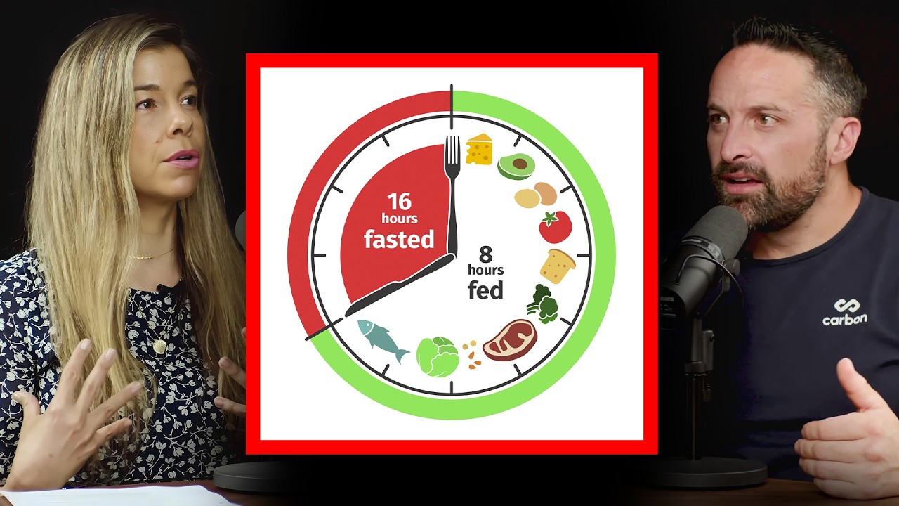
In this clip, Drs. Rhonda Patrick and Layne Norton discuss the benefits, limits, and longevity impacts of time-restricted eating and calorie restriction.
In this clip, Dr. Martin Gibala outlines the relationship between exercise types, mitochondrial growth, and their combined effect on fat metabolism.
-
Dr. Rhonda Patrick discusses child nutrition, osteoporosis, cholesterol-lowering supplements, her skincare routine, and fasting versus prioritizing protein.
-
In this clip, Drs. Rhonda Patrick and Layne Norton discuss the benefits, limits, and longevity impacts of time-restricted eating and calorie restriction.
-
In this clip, Dr. Martin Gibala outlines the relationship between exercise types, mitochondrial growth, and their combined effect on fat metabolism.
-
Autophagy facilitates ATP release from dying cells, triggering immunosurveillance by attracting myeloid cells through purinergic receptors.
-
In this clip, Dr. Mark Mattson describes the importance of transient, short-term, mild energetic stress to maintain healthy cells.
-
In this clip, Dr. Mark Mattson predicts that intermittent fasting benefits both overweight and healthy weight people.
-
Coffee consumption is associated with protection from several chronic diseases. Coffee's beneficial effects may stem from its ability to induce autophagy; ho...
-
Aliquot #15: Autophagy: A cellular recycling program with implications for health and longevity PremiumAutophagy, a type of cellular recycling program, influences many aspects of health and disease, including cancer, neurodegenerative disease, and aging. Nutri...
-
In this clip, Dr. Satchin Panda explains the rationale behind the metabolic benefits associated with time-restricted eating.
-
Q&A Mashup #3: Fasting PremiumThis episode of the aliquot features a collection of clips covering a range of topics related to fasting - both intermittent and prolonged. Specific topics ...
-
Dr. Rhonda Patrick answers audience questions on various health, nutrition, and science topics in this Q&A session.
-
Rhonda Vitamin D Exercise Sleep Omega-3 Fasting Pregnancy ADHD Muscle Autophagy Resveratrol Sulforaphane Metformin Supplements Ketogenic Diet Wearable TechnologyDr. Rhonda Patrick answers audience questions on various health, nutrition, and science topics in this Q&A session.
-
Resveratrol has positive impact on brain health & Alzheimer’s disease prevention | David Sinclair ClipIn this clip, Dr. David Sinclair and Dr. Rhonda Patrick discuss the brain health benefits associated with resveratrol.
-
Dr. Longo discusses research indicating that the fasting-mimicking diet may hold promise for autoimmune diseases, especially multiple sclerosis.
-
Dr. Valter Longo discusses how cellular repair mechanisms may have evolved during times of fasting, but are latent in times of food abundance.
-
Dr. Rhonda Patrick describes some of the different types of fasting and identifies the advantages associated with each.
-
Dr. Valter Longo describes how prolonged fasting and subsequent refeeding, sends signals to the body to systematically shrink and rebuild organ systems.
-
Dr. Valter Longo explains the benefits of a prolonged fast.
-
Mitophagy: a signal for mitochondrial biogenesis, metabolic adaptation, and cellular differentiation ClipDr. Guido Kroemer describes mitophagy, a cellular signal for mitochondrial biogenesis, and a key player in other physiological processes.
-
How the cell prioritizes and targets organelles and protein aggregates for degradation by autophagy ClipDr. Guido Kroemer describes the cellular process of autophagy and what factors trigger its activation.
-
Increasing general autophagy as a treatment for neurodegenerative diseases like Parkinson's disease ClipDr. Guido Kroemer describes how mitophagy contributes to the pathophysiology of neurodegenerative disease and how autophagy might mitigate these processes.
-
Fasting associated with sickness behavior critical to surviving bacterial infection | Guido Kroemer ClipDr. Guido Kroemer describes how fasting associated with sickness behavior is critical to surviving infection.
-
Dr. Guido Kroemer describes the complex relationship that exists between cancer cells, the immune system, and autophagy.
-
Deprivation of nutrients (starvation) and other signals that play a key role in inducing autophagy ClipDr. Guido Kroemer discusses changes that occur within cells that signal the need to induce autophagy.
-
Dr. Guido Kroemer describes the anti-diabetes and anti-obesity effects of exercise-induced autophagy.
-
Dr. Guido Kroemer describes the interplay between protein deacetylation and autophagy.
-
Dr. Guido Kroemer describes how intermittent fasting recapitulates the life-extension benefits of caloric restriction without some of the drawbacks.
-
Dr. Guido Kroemer discusses the autophagy-inducing qualities of coffee.
-
Autophagy expert: Dr. Guido Kroemer's personal take on fasting, eating and other lifestyle habits ClipDr. Guido Kroemer describes the autophagy-inducing behaviors he practices as part of his healthy lifestyle.
-
Caloric Restriction Mimetics in Aging, Improved Cancer Chemotherapy, Autophagy Anti-Obesity Effect ClipDr. Guido Kroemer describes the autophagy-inducing effects of calorie restriction mimetics such as spermidine and resveratrol.
-
Dr. Guido Kroemer describes the spectacular biological phenomenon known as autophagy.
-
Dr. Guido Kroemer describes the current state of knowledge about the minimum amount of time required for fasting-induced autophagy in humans.
-
Dr. Dale Bredesen describes his novel dietary protocol, the Ketoflex 12/3, and how it improves cognitive function.
-
Dr. Ruth Patterson describes how eating following the body's circadian clock enables the body to focus on repair rather than on digestion.
-
This episode features a Q&A session on fasting-related topics with Dr. Rhonda Patrick and Mike Maser of Zero Fasting Tracker.
-
Rhonda Vitamin D Brain Alzheimer's Gut Microbiome Sleep Fasting Autophagy Sauna Vegetarian Weight Loss Supplements Ketogenic Diet Wearable Technology Blood TestDr. Rhonda Patrick answers audience questions on various health, nutrition, and science topics in this Q&A session.
-
Dr. Valter Longo on Resetting Autoimmunity and Rejuvenating Systems with Prolonged Fasting & the FMDFasting Cancer Obesity Aging Heart Disease Diabetes Insulin Resistance Inflammation Stem Cells Immune System Tissue Repair Autophagy Apoptosis Insulin AutoimmunityDr. Valter Longo discusses fasting as a means to treat or prevent age-related diseases such as cancer, Alzheimer’s disease, and others.
-
Epigenetics Vitamin D Nutrition Exercise Aging Metabolism Sleep Diabetes Telomeres DNA Damage Stem Cells Stress Melatonin Vitamin E Genetics 23andMe Heat Stress Autophagy Autism Folate Sauna AntioxidantNutrigenomics examines how genetic variation influences micronutrient absorption and metabolism, and the biological consequences of these interactions.
-
Rhonda Nutrition Alzheimer's Cancer Gut Fasting Circadian Rhythm Pregnancy Vaccine Autophagy Sulforaphane Time-Restricted Eating Breast Milk Supplements Ketogenic DietDr. Rhonda Patrick answers audience questions on various health, nutrition, and science topics in this Q&A session.
-
Aging Nutrition Cancer Podcast Fasting Multiple Sclerosis Autophagy Video Triglycerides Fatty Liver Time-Restricted Eating ProteinDr. Guido Kroemer discusses immunology, cancer biology, calorie-restriction mimetics, aging, and autophagy.
-
Nutrition Vitamin D Metabolism Diabetes Telomeres Omega-3 Inflammation Depression DNA Damage Stem Cells Micronutrients Mitochondria Autophagy Autism Schizophrenia Resveratrol Sulforaphane Insulin Blue LightDr. Rhonda Patrick makes her fifth appearance on the Joe Rogan Experience.
-
Dr. Valter Longo discusses the role of fasting and the fasting-mimicking diet in longevity, cancer & multiple sclerosis.
-
Dr. Rhonda Patrick is on The Tim Ferriss Show in episode #12 entitled "Rhonda Patrick on Life Extension, Performance, and More".
Topic Pages
-
Autophagy
Autophagy is a conserved catabolic pathway where double-membrane autophagosomes deliver cytoplasmic cargo to lysosomes for degradation.
-
Fasting
Fasting-induced nutrient deprivation inhibits mTORC1, activates the ULK1 complex, thereby promoting macroautophagy-mediated intracellular component recycling.
-
Rapamycin
Rapamycin induces autophagy by allosterically inhibiting mTORC1, relieving its suppression of ULK1-initiated autophagosome biogenesis.
News & Publications
-
As brain cells age, their cleanup systems lose efficiency, allowing damaged parts and sticky proteins to build up. To address this, a new study tried to restore guanosine triphosphate (GTP), which fuels cargo transport and recycling inside cells, to see if this could slow or even reverse the decline in aging neurons.
In neurons from healthy older mice and from a strain that develops Alzheimer-like changes, researchers tested whether a 24-hour treatment could restore GTP levels and improve waste processing. The treatment used two bioactive compounds: nicotinamide, a vitamin B3 derivative, and epigallocatechin gallate (EGCG), a green tea compound.
The experiments revealed clear age-related declines as well as improvements after treatment:
- Aging neurons had lower GTP levels, including less free GTP near mitochondria. This decline appeared earlier in the Alzheimer-like strain.
- Treatment restored GTP to levels seen in young neurons.
- Signs of stalled transport inside cells decreased, suggesting cargo was moving to its destination more effectively.
- Amyloid-beta protein levels decreased inside neurons. Normally present in the brain, this protein can accumulate and form toxic clumps in Alzheimer's disease, damaging brain cells.
- When recycling pathways (autophagy) were blocked, GTP increased. When recycling was stimulated, GTP decreased. This confirmed that cleanup consumes significant GTP.
Two key cleanup routes in brain cells depend on GTP:
- Endocytosis brings materials into the cell for sorting or breakdown.
- Autophagy dismantles worn or damaged components and delivers them to the lysosome, a small acidic compartment that digests waste.
Both routes rely on moving materials inside the cell using tiny carriers that deliver their cargo to specific locations. This transport is directed by GTP-binding "switch" proteins, which turn on to move a carrier to the correct location and turn off once the delivery is complete. In older neurons, reduced GTP levels may slow these steps. Nicotinamide may help restore the cell's energy capacity, while EGCG may activate protective genes through the Nrf2 pathway, helping maintain the conditions needed for transport and recycling to function properly.
Conclusion:
The study shows that aging neurons had lower GTP and signs of slowed waste processing. Treatment with nicotinamide and EGCG restored GTP and improved cleanup markers. These results point to an energy bottleneck in cellular cleanup that can be relieved under controlled settings. However, the findings do not yet address brain function, long-term safety, or effectiveness in animals or humans. Learn more about how you can support your brain's ability to clear waste products in this episode. -
Chronic stress inhibits autophagy—the brain's recycling system—but restoring its functionality yields rapid antidepressant effects. pubmed.ncbi.nlm.nih.gov
In small doses, stress can sharpen focus and improve resilience, but chronic stress gradually erodes emotional stability, increasing the risk of major depressive disorder. A recent study found that autophagy—the brain’s recycling and housekeeping system—helps maintain emotional stability by removing old or damaged proteins.
Researchers explored how short-term and long-term stress influenced autophagy in mice and investigated whether antidepressant drugs could restore this process. Employing genetic techniques, the researchers selectively inhibited or enhanced autophagy in a region of the brain called the lateral habenula and then monitored how the animals reacted to stress.
They found that acute stress activated autophagy, while chronic stress inhibited it. When autophagy ceased functioning properly, stress-related behaviors increased. However, restoring autophagy—even briefly—produced rapid antidepressant-like effects. Drugs commonly used to treat depression also reactivated autophagy in this brain region. Additional experiments indicated that autophagy helps regulate brain cell activity by breaking down excess glutamate receptors, which are often overactive in depression.
These findings suggest that disrupted autophagy in the lateral habenula plays a central role in how chronic stress contributes to depression. Learn more about autophagy in this episode featuring Dr. Guido Kroemer.
-
Resistance exercise activates muscles' cellular "housekeeping" processes, ridding cells of harmful waste products. pubmed.ncbi.nlm.nih.gov
Cellular processes are messy. They produce copious amounts of harmful waste products and damaged parts that can accumulate inside the cell, creating havoc and even cell death. A recent study found that resistance training activates critical cellular cleanup processes in muscles, facilitating waste disposal and supporting muscle cell health.
The study had two phases: an acute exercise phase with intense workouts to trigger an immediate muscle response and a six-week adaptation phase to see how muscles adjust to repeated mechanical stress. In the acute phase, participants performed resistance exercises, including leg extensions, leg presses, and drop jumps, using heavy weights and incorporating eccentric movements. During the adaptation phase, they performed the exercises at a lower intensity twice a week, allowing the muscles to adapt to the consistent workload gradually. Researchers took muscle samples before and after these sessions at the start and end of the study, analyzing changes in critical proteins and focusing on a cellular protein called BAG3.
BAG3 facilitates a cellular cleanup process called chaperone-assisted selective autophagy, or CASA. Unlike ordinary autophagy, which generally recycles various cell parts, CASA specifically targets and breaks down damaged or misfolded proteins with the help of unique proteins called chaperones. BAG3 is one of these chaperones. Under resting conditions, two phosphorous molecules tether BAG3 in place, keeping it inactive.
However, the researchers found that when cells experienced mechanical stress, the phosphorous molecules released BAG3, activating it and allowing it to perform its chaperoning job. Notably, BAG3 release was essential for eliminating damaged mitochondria—a process called mitophagy—in skeletal muscles.
These findings suggest resistance exercise supports cellular health and mitophagy by boosting in-house cleanup processes. Learn more about mitophagy in this clip featuring Dr. Guido Kroemer.
-
Time-restricted eating activates genes involved in metabolism and autophagy. www.sciencedaily.com
Time-restricted eating influences the activation of roughly 70 percent of all genes in mice, a new study shows. Mice that ate on a time-restricted schedule had fewer active genes involved in inflammation and oxidative stress and more active genes involved in metabolism and autophagy – a cellular defense mechanism.
Researchers fed two groups of mice a Western-style diet, which is high in fat and sugars, for seven weeks. One group was allowed to eat whenever they chose to, but the other group was allowed to eat only during a nine-hour window each day. At the end of the seven-week intervention, the researchers analyzed gene activity in the animals' tissues at different times of the day.
They found that time-restricted eating altered the activity of more than 80 percent of genes involved in protein synthesis, folding, and maintenance. They also found that time-restricted eating altered amino acid, fat, and glucose metabolism and re-aligned the circadian rhythms of the animals' organs.
These findings suggest that time-restricted eating influences gene activity in mice. If the findings translate to humans, they could have far-reaching implications for chronic metabolic disorders, neurodegenerative diseases, cancer, and other diseases. Learn more about the health benefits of time-restricted eating in this episode featuring Dr. Satchin Panda, the senior investigator for this study.
-
Eating twice a day, without restricting calories, may prevent metabolic syndrome.
Metabolic syndrome is a constellation of medical conditions that includes high blood pressure, high blood glucose, unhealthy cholesterol levels, and excess abdominal fat. Having metabolic syndrome increases a person’s risk of heart disease, stroke, and diabetes. Findings from a 2017 study suggest that eating two meals per day, without caloric restriction, induces inter-meal autophagy, potentially preventing metabolic syndrome.
Calorie restriction is the practice of long-term reduced dietary intake, typically characterized by a 20 to 50 percent decrease in caloric intake below habitual levels, without malnutrition or deprivation of essential nutrients. Evidence indicates that calorie restriction induces autophagy; however, adherence to the practice is challenging and often not sustainable.
Autophagy is a highly conserved adaptive response to stress that involves the sequestration and subsequent destruction of protein aggregates, pathogens, and damaged or dysfunctional cellular components. The primary goal of autophagy is to allow cells to adapt to changing conditions and external stressors, including nutrient scarcity.
The investigators examined the effects of different dietary patterns on autophagy in mice. They fed one group of mice two distinct meals per day (morning and evening), with each meal providing an equal number of calories. They fed another group of mice the same number of calories, but the mice were allowed to eat anytime during a 24-hour period. They monitored the animals' bodyweight and body composition and measured markers of autophagy and metabolic function.
They found that the two groups of mice weighed about the same at the end of the 16-month intervention, but mice that ate two distinct meals per day had less bodyfat and more muscle mass than those that ate freely all day. Mice that ate twice daily also exhibited increased expression of autophagy-related genes; browning of white adipose tissue (a phenomenon associated with improved metabolic function in mice); increased the expression of M2 macrophages, which exert anti-inflammatory properties; and improved aspects of metabolism.
These findings suggest that eating just two meals per day, without restricting total calories, induces autophagy between meals and improves aspects of metabolism. Watch this episode in which Dr. Guido Kroemer describe the effects of calorie restriction on autophagy.
Read our overview articles on autophagy and calorie restriction to learn more.
-
Spermidine supplementation improves memory performance in older adults with subjective cognitive decline. pubmed.ncbi.nlm.nih.gov
Spermidine is a polyamine compound that may increase health span due to its ability to induce autophagy, the process by which the body removes damaged and dysfunctional cells. In animal models, spermidine supplementation has been shown to prevent memory loss. Findings from a recent report detail the first experiment exploring the effects of spermidine supplementation on memory in older adults without dementia.
Episodic memory, which records specific events, situations, and experiences, declines with age, but this loss may be impeded by certain lifestyle interventions, such as caloric restriction. The effects of spermidine in the body mimic caloric restriction, making it a promising therapy for the reversal of memory loss. Previous research demonstrates the ability of spermidine supplementation to restore memory performance in fruit flies; however, the effects of spermidine supplementation on memory performance in humans are unknown.
The authors recruited 30 adults (aged 60 to 80 years) with subjective cognitive decline, a condition associated with objective cognitive decline and Alzheimer’s disease. They assigned half of the participants to consume a capsule containing 750 milligrams of a spermidine-rich plant extract containing 1.2 milligrams of spermidine daily for three months, while the other half consumed a placebo supplement. Participants completed memory assessments and other cognitive testing before and after the supplement period.
Participants consuming the spermidine supplement had moderately enhanced memory performance after three months compared to those who took the placebo. In particular, permidine supplementation enhanced mnemonic discrimination, the ability to differentiate between new and previously encountered items. There was no difference in other cognitive functions between groups.
The authors concluded that spermidine supplementation may be an effective treatment for slowing cognitive decline in older adults with subjective cognitive impairment. They noted that this was a small pilot trial and that larger clinical trials are needed to expand on these results.
-
Spermidine improves cognitive function in humans. www.cell.com
Age-related changes in the brain drive cognitive impairment, which in turn alters memory formation and promotes mitochondrial dysfunction. A recent study demonstrates that spermidine improves cognitive function in humans.
Spermidine is a dietary compound found in a number of foods, including wheat germ, cauliflower, broccoli, mushrooms, amaranth, a variety of cheeses (especially aged cheeses), and soybean products, such as natto. First identified in semen, spermidine demonstrates anti-aging properties in animal studies, likely due to its capacity to induce autophagy – the process by which the body clears dead and damaged cells. Human studies have demonstrated an association between dietary spermidine intake and increased survival..
The authors of the study investigated the effects of spermidine supplementation in various models of aging. They fed aged mice spermidine in their drinking water and measured the compound’s appearance in the animals' brains. The compound appeared in brain tissue within one week of administration and continued to accrue to roughly one-third of bloodstream levels. Spermidine altered protein synthesis in the animals' brains via its actions on hypusine, an amino acid that is essential for the function of ELF5A, a protein that plays roles in mammalian protein production. Mice that received spermidine exhibited improvements in spontaneous behavior, exploration, maze completion, and other measures of cognitive function.
The authors also fed fruit flies spermidine and found that the compound improved mitochondrial function and reduced age-induced memory impairments. These improvements were mediated by Atg7, PINK, and parkin – proteins that play key roles in autophagy and mitophagy.
Then the authors reviewed epidemiological data from food frequency questionnaires and cognitive tests completed by participants in the Bruneck Study, a prospective cohort study of adults living in Bruneck, Italy. They found that higher intake of spermidine-rich foods was associated with greater cognitive function in aging.
These findings suggest that dietary spermidine exerts neuroprotective effects in several model of aging, likely due to the compound’s effects on mitochondrial health. Dietary interventions that promote spermidine-rich foods may be useful in slowing cognitive decline in humans.
-
The circadian clock coordinates behavioral and circadian cues with the availability and utilization of nutrients. Proteasomal degradation of clock repressors, e.g., cryptochrome (CRY)1 maintains periodicity of the clock. Whether autophagy, a quality control pathway, degrades circadian proteins remains unknown. Here we show that circadian proteins BMAL1, CLOCK, REV-ERB, and CRY1 are lysosomal targets, and that α macroautophagy (hereafter autophagy) specifically degrades CRY1. Autophagic degradation of CRY1, an inhibitor of gluconeogenesis, occurs in a diurnal window when rodents rely on gluconeogenesis, suggesting that degradation of CRY1 is time-imprinted to maintenance of blood glucose levels. CRY1 contains several light chain 3 (LC3)-interacting region (LIR) motifs, which facilitate the interaction of cargo proteins to the autophagosome marker LC3. Using mutational analyses, we identified two distinct LIRs on CRY1 that exert circadian control over blood glucose levels by regulating CRY1 degradation, revealing CRY1 LIRs as potential targets in regulation of glucose metabolism.
Toledo, Miriam and Tarabra, Elena and Batista-Gonzalez, Ana and Merlo, Paola and Feng, Daorong and Sarparanta, Jaakko and Botrè, Francesco and Pessin, Jeffrey E. and Singh, Rajat, Autophagy Regulates the Liver Clock and Glucose Metabolism by Degrading CRY1 (2018). Available at SSRN: https://ssrn.com/abstract=3155564 or http://dx.doi.org/10.2139/ssrn.3155564
-
Hair Regeneration by Small Molecules That Activate Autophagy papers.ssrn.com
Hair plays important roles, ranging from the conservation of body heat to the preservation of psychological well-being. Hair loss or alopecia affects millions worldwide and can occur because of aging, hormonal dysfunction, autoimmunity, or as a side effect of cancer treatment. Methods that can be used to regrow hair are highly sought after, but lacking. Here we report that hair regeneration can be stimulated by small molecules that activate autophagy, including the longevity metabolites α-ketoglutarate and α-ketobutyrate, and the prescription drugs rapamycin and metformin which impinge on TOR and AMPK signaling.
Chai, Min and Jiang, Meisheng and Vergnes, Laurent and Fu, Xudong and de Barros, Stéphanie C. and Jiao, Jing and Herschman, Harvey R. and Crooks, Gay M. and Reue, Karen and Huang, Jing, Hair Regeneration by Small Molecules That Activate Autophagy (2018). Available at SSRN: https://ssrn.com/abstract=3188356 or http://dx.doi.org/10.2139/ssrn.3188356
-
Acid Suspends the Circadian Clock in Hypoxia through Inhibition of mTOR. - PubMed - NCBI www.ncbi.nlm.nih.gov
Citation: Cell. 2018 Jun 28;174(1):72-87.e32. doi: 10.1016/j.cell.2018.05.009. Epub 2018 May 31.
Abstract Recent reports indicate that hypoxia influences the circadian clock through the transcriptional activities of hypoxia-inducible factors (HIFs) at clock genes. Unexpectedly, we uncover a profound disruption of the circadian clock and diurnal transcriptome when hypoxic cells are permitted to acidify to recapitulate the tumor microenvironment. Buffering against acidification or inhibiting lactic acid production fully rescues circadian oscillation. Acidification of several human and murine cell lines, as well as primary murine T cells, suppresses mechanistic target of rapamycin complex 1 (mTORC1) signaling, a key regulator of translation in response to metabolic status. We find that acid drives peripheral redistribution of normally perinuclear lysosomes away from perinuclear RHEB, thereby inhibiting the activity of lysosome-bound mTOR. Restoring mTORC1 signaling and the translation it governs rescues clock oscillation. Our findings thus reveal a model in which acid produced during the cellular metabolic response to hypoxia suppresses the circadian clock through diminished translation of clock constituents.
-
Disruption of the beclin 1–BCL2 autophagy regulatory complex promotes longevity in mice www.nature.com
[Abstract]
Autophagy increases the lifespan of model organisms; however, its role in promoting mammalian longevity is less well-established. Here we report lifespan and healthspan extension in a mouse model with increased basal autophagy. To determine the effects of constitutively increased autophagy on mammalian health, we generated targeted mutant mice with a Phe121Ala mutation in beclin 1 (Becn1F121A/F121A) that decreases its interaction with the negative regulator BCL2. We demonstrate that the interaction between beclin 1 and BCL2 is disrupted in several tissues in Becn1F121A/F121A knock-in mice in association with higher levels of basal autophagic flux.
Compared to wild-type littermates, the lifespan of both male and female knock-in mice is significantly increased. The healthspan of the knock-in mice also improves, as phenotypes such as age-related renal and cardiac pathological changes and spontaneous tumorigenesis are diminished. Moreover, mice deficient in the anti-ageing protein klotho3 have increased beclin 1 and BCL2 interaction and decreased autophagy. These phenotypes, along with premature lethality and infertility, are escued by the beclin 1(F121A) mutation. Together, our data demonstrate that disruption of the beclin 1–BCL2 complex is an effective mechanism to increase autophagy, prevent premature ageing, improve healthspan and promote longevity in mammals.
-
Fasting-like diet kills autoimmune cells & replaced them with healthy cells & improved symptoms in MS patients. www.sciencedaily.com
This fasting-like diet also promotes regeneration of the myelin in mice with multiple sclerosis. In human patients with multiple sclerosis, the fasting-like diet led to improvements in symptoms if followed by a Mediterranean diet or a ketogenic diet.
Here is the fasting-like diet that humans were given: Day 1 – pre-fasting followed by Day 2-8 – very low calorie diet. Day 1-prefasting consists of an 800 kcal (about 40% of normal caloric intake similar to mouse Day1 FMD) monodiet (fruit, rice, or potatoes) by preference of individuals. On the following day patients were recommended to use an oral laxative, Natrium Sulfuricum (20-40 g). FMD consisted of 100 ml vegetable broth or vegetable juice with 1tablespoon of linseed oil 3 times daily, plus additional calorie-free liquids. The daily calorie intake was predefined with 200 – 350 kcal (10-18% of normal caloric intake similar to mouse Day 2-3 FMD). Patients were advised to drink 2-3 L of unsweetened fluids each day (water, and herbal teas) and to use an enema if tolerated. After the 7-day fasting period solid foods were stepwise reintroduced for three days, starting with a steamed apple at day 8. After the fasting and refeeding period a normocaloric, plant-based Mediterranean diet was maintained until study end.